Chapter: Human Neuroanatomy(Fundamental and Clinical): Cranial Nerve Nuclei
Cranial Nerve Nuclei
Cranial Nerve Nuclei
The seven functional components to which the fibres of a cranial nerve may belong are as follows.
(1) Somatic efferent. (2) General visceral efferent. (3) Special visceral efferent. (4) Generalsomatic afferent. (5) Special somatic afferent. (6) General visceral afferent. (7) Special visceral afferent.
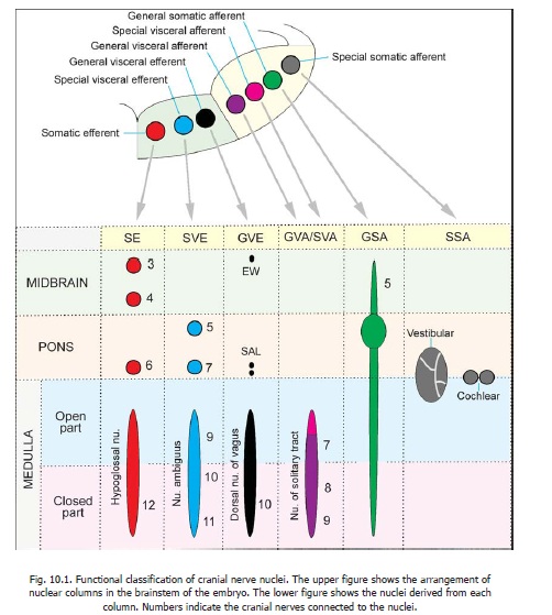
Each functional component has its own nuclei of origin (in the case of efferent fibres) or of termination (in the case of afferent fibres). In the embryo the nuclei related to the various components are arranged in vertical rows (or columns) in a definite sequence in the grey matter related to the floor of the fourth ventricle (Fig. 10.1). The sequence is easily remembered if the following facts are kept in mind.

a. Each half of the floor of the ventricle is divided into a medial part and a lateral part by the sulcuslimitans. Efferent nuclei lie in the medial part (called the basal lamina) and afferent nuclei inthe lateral part (called the alar lamina).
b. In each part (medial or lateral) visceral nuclei lie nearer the sulcus limitans than somaticnuclei.
c. Within each category (e.g., visceral efferents, somatic afferents etc.) the general nucleus lies nearer the sulcus limitans than thespecial nucleus.
Thus in proceeding laterally from the midline the sequence of nuclear columns is as follows.
1. Somatic efferent (SE). This column is not subdivided into general and special parts.
2. Special visceral (or branchial) efferent (SVE).
3. General visceral efferent (GVE).
4. General visceral afferent (GVA).
5. Special visceral afferent (SVA).
6. General somatic afferent (GSA).
7. Special somatic afferent (SSA).
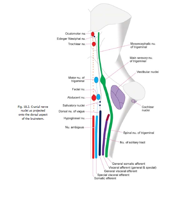
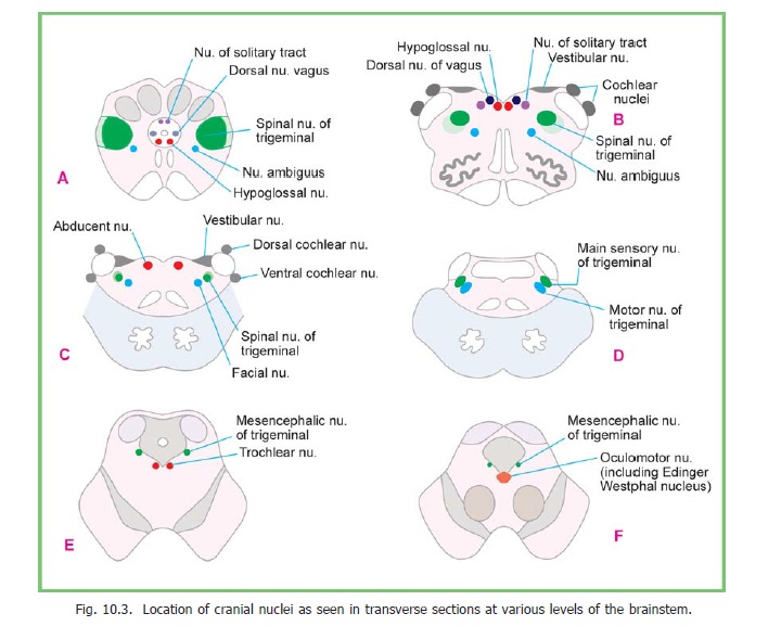
As development proceeds parts of these columns disappear so that each of them no longer extends the whole length of the brainstem, but is represented by one or more discrete nuclei. These nuclei are shown schematically in the lower half of Fig. 10.1. Some nuclei retain their original positions in relation to the floor of the fourth ventricle, but some others migrate deeper into the brainstem. The position of the nuclei relative to the posterior surface of the brainstem is illustrated in Fig. 10.2. The positions of the nuclei as seen in transverse sections of the brainstem are shown in Fig. 10.3.

In the description that follows the nuclei of the third to twelfth cranial nerves are considered as they are located in the brainstem. The third and fourth nerves belong to the midbrain; the fifth, sixth, seventh (and part of eighth) to the pons; and the remaining to the medulla.

Related Topics