Chapter: Human Neuroanatomy(Fundamental and Clinical): Cranial Nerve Nuclei
General and Special Somatic Afferent Nuclei - Cranial Nerve Nuclei
General Somatic Afferent Nuclei
The general somatic afferent column is represented by the sensory nuclei of the trigeminal nerve. These are as follows.
1. Themain(orsuperior)sensory nucleuslies in the upper part of the pons, in the lateral partof the reticular formation. It lies lateral to the motor nucleus of the trigeminal (Figs 10.1, 10.2 and 10.3D). It is mainly concerned in mediation of proprioceptive impulses, touch and pressure (from the region to which the trigeminal nerve is distributed).
2. Thespinal nucleusextends from the main nucleus down into the medulla (Figs 10.1, 10.2 and10.3 A,B,C) and into the upper two segments of the spinal cord. Its lower end is continuous with the substantia gelatinosa of the cord. In addition to fibres of the trigeminal nerve, the nucleus also receives general somatic sensations carried by the facial, glossopharyngeal and vagus nerves.The spinal nucleus is concerned mainly with the mediation of pain and thermal sensibility. Different parts of the nucleus correspond to different areas innervated. The spinal nucleus is divisible (cranio-caudally) into three sub-nuclei, oralis, interpolaris, and caudalis.
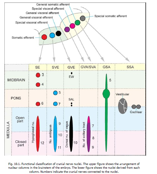
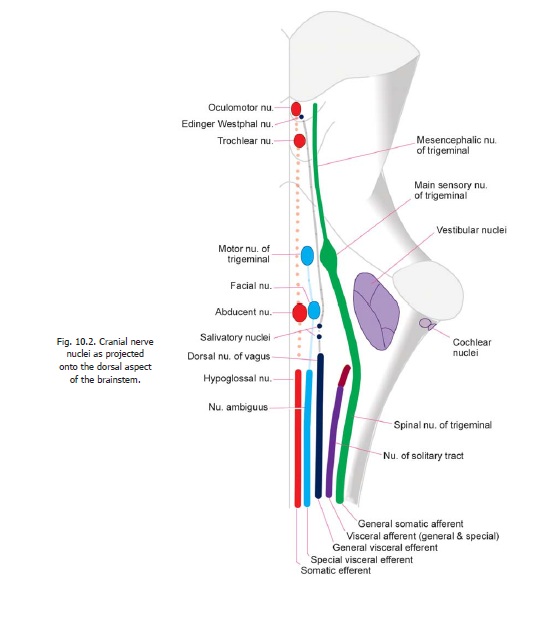
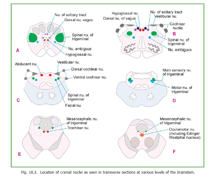
3. Themesencephalic nucleusof the trigeminal nerve extends cranially from the upper end ofthe main sensory nucleus into the midbrain. Here it lies in the central grey matter lateral to the aqueduct (Fig. 10.3 E,F). Functionally, this nucleus appears to be similar to sensory ganglia of cranial nerves, and to the spinal ganglia, rather than to afferent nuclei. The neurons in it are unipolar. The peripheral processes of these neurons are believed to carry proprioceptive impulses from muscles of mastication, and possibly also from muscles of the eyeballs, face tongue and teeth. The central processes of the neurons in the nucleus probably end in the main sensory nucleus of the trigeminal nerve.
The mesencephalic nucleus is the centre for the jaw jerk
Special Somatic Afferent Nuclei
These are the cochlear and vestibular nuclei.
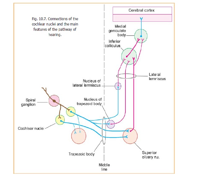
The cochlear nuclei are two in number, dorsal and ventral. They are placed dorsal and ventral, respectively, to the inferior cerebellar peduncle (Fig. 10.3 B,C) at the level of the junction of the pons and medulla. The two nuclei are continuous, being separated only by a layer of nerve fibres. Their connections are shown in Fig. 10.7.
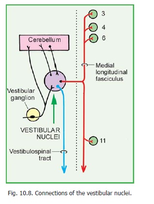
The vestibular nuclei lie in the grey matter underlying the lateral part of the floor of the fourth ventricle (Fig.10.3 B,C). They lie partly in the medulla and partly in the pons. Four distinct nuclei are recognized: these are medial, lateral, inferior, and superior. The lateral nucleus is also called Dieter’snucleus. The connections of the vestibular nuclei are shown in Fig.10.8.
Related Topics