Chapter: 11th Zoology : Chapter 12 : Basic Medical Instruments and Techniques
Imaging Medical Instruments
Imaging Instruments
EEG – Electroencephalogram
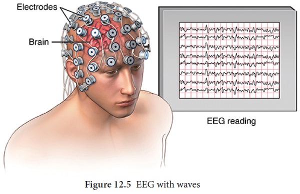
EEG is a test to evaluate the electrical activities of the brain (Figure12.5). Brain cells communicate with each other through electrical impulses. An EEG tracks and records the brain wave patterns. It is a graphical recording of the electrical activities of the cortical and sub cortical regions of the brain. It is recorded by placing the surface electrodes on the scalp region. The electrodes analyse the electrical impulses in the brain and send signals to a computer which records the results. In 1929 Germen scientist Hans Berger was the first to analyse the EEG. Hence, EEG is also known as “Berger Wave”. The electrical activity recorded by EEG may have synchronised or desynchronised waves. It has four frequency waves/ rhythms namely alpha, beta, delta and theta waves.
Clinical significance of EEG
1.
EEG provides a means to study the functioning of
the brain and its coordination with other parts of the body.
2.
It is useful in diagnosis of neurological and sleep
disorders
3.
EEG has proved to be a useful diagnostic tool in
cases of serious head injuries, brain tumours and cerebral infections.
4.
It also helps to find the diseases like epilepsy
and various degenerative disease of the nervous system.
5.
EEG is useful in assessing patients with suspected
brain death.
X-rays
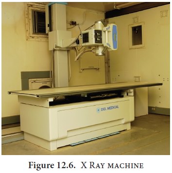
Radiography is the use of X-rays
to visualize the internal structures of a patient (Figure12.6). X-Rays are a form
of electromagnetic radiation, produced by an X-ray tube. The X-rays are passed
through the body and captured behind the patient by a detector; film sensitive
to x-rays or a digital detector. There is variation in absorption of the X-rays
by different tissues within the body. Dense bones absorb more radiation, while
soft tissue allows more rays to pass through. This variation produces contrast
image within the image to give a 2D representation of all the structures. It is
invasive. It is also available as a portable X ray unit. It is less costly when
compared to the other imaging units like MRI or CT.
Clinical significance
1.
X- ray imaging is used for diagnosing the disease
of the heart, lungs and fractures of bones and joints
2.
It is also used to visualise hollow organs and
blood vessels by filling them with certain chemical formulations containing
barium and iodine
3.
Dental radiography is used in diagnosis of oral
problems
4.
Mammography is a special type of X-ray imaging to
create detail images of the breast tissues
5.
Fluoroscopy for real time images
6.
X-rays are used in radiation therapy to shrink
cancerous tumours
Ultrasound imaging
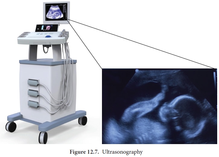
Ultrasound literally means sound beyond the range
of human hearing. Ultrasound waves are produced by a physical phenomenon known
as Piezo-electric effect. When an electric potential is applied to certain
crystals for example: Lead zirconate, they become excited, vibrate and produce
ultrasound. When the ultrasound waves are introduced through homogenous tissue,
they pass unimpeded until they meet another tissue or organ. A part or whole of
the ultrasound wave is reflected and received back by the same crystal and is
converted into an electrical signal. This signal denoting reflecting interface
is shown on the oscilloscope screen as a deflection from the base line
(Figure12.7).
Clinical significance
1. Ultrasound
waves are used to image the foetus at different stages of pregnancy to study
the progress of the developing foetus.
2. They are
used to hear foetal heart sound, blood flow, etc.
3. Used in
echocardiography to diagnose the damages in heart.
4. Used for
diagnosis of tumours, gall stones, kidney stones, obstructions in the genital
tracts.
Computed Tomographic (CT) Scanning
Computed tomography is originally known as computed
axial tomography (CAT or CT Scan). The word tomography is derived from the
Greek word tomos means slice and graphe means to
write. It is a medical imaging technology employing tomography, were digital
geometry processing is used to generate a three dimensional image of the
internals of an object from a large series of two dimensional X ray images
taken around a single axis of rotation (Figure12.8).
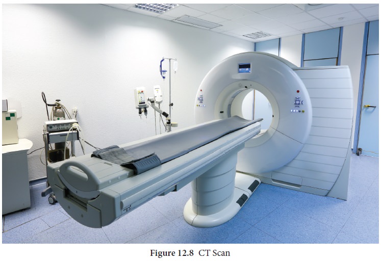
CT produces volumes of data which can be
manipulated through a process known as windowing in order to demonstrate
various structures based on their ability to block the X ray beam.
Clinical significance
•
Gives a clear image of bone, soft tissues and blood
vessels
•
Helps in the diagnosis of injuries of the inner
ears and sinuses
•
To
detect cancer, heart and
lung Disorders
•For diagnosis of spinal problems and skeletal
injuries
•
Helps to measure bone mineral density
•
To detect stroke causing clots and hemorrhage in
the brain.
Positron Emission Tomographic Scanning (PET)
PET is also computerized imaging technique unlike
CT. Positron emission tomography (PET) is a nuclear medicine procedure based on
the measurement of positron emission from radiolabelled tracer molecules. These
radiotracers allow biologic processes to be measured and whole body images to
be obtained which demonstrates sites of radiotracer accumulation. A PET image
gives quantitative regional information on the metabolic and physiological
processes. PET uses positron emitting radio isotopes (11C 13N 15O 18F) which
are generated by the cyclotron. The most common radiotracer in use today is
18F- fluorodeoxyglucose (18F-FDG) which is a radio labelled sugar
(glucose) molecule. These atoms are then incorporated by chemical methods into
biological molecules like glucose, amino acids and ammonia. These positron
emitting compounds are then injected in very small amounts into or inhaled by
experimental animals or human subjects. The three dimensional distribution of
the labeled trace is then probed by powerful PET cameras and the images are
reconstructed by a computer. The quantitative interpretation of the image is
done by varying mathematical models. They deal with the process of uptake and
metabolism of the radioisotope.
Clinical significance
PET imaging is effectively used in the measurement
of regional cerebral blood volume, blood flow, metabolic rates for glucose and
oxygen in humans.
NMRI-Magnetic Resonance Imaging
Magnetic resonance imaging (MRI) is a non-invasive
medical test that physicians use to diagnose medical conditions. Unlike
conventional X-ray examinations and computed tomography (CT) scans,MRI does not
utilize ionizing radiation. MRI uses a powerful magnetic field, radio frequency
pulses and a computer to produce detailed pictures of organs, soft tissues,
bone and virtually all other internal body structures. The radio frequency
pulses re-align hydrogen atoms that naturally exist within the body while the
patient is in the scanner without causing any chemical changes in the tissues.
As the hydrogen atoms return to their usual alignment, they emit different
amounts of energy that vary according to the type of body tissue from which
they come. The MR scanner captures this energy and creates a picture of the
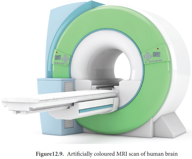
tissues scanned based on this information (Figure12.9).
The magnetic field is produced by passing an
electric current through wire coils in most MRI units. Other coils, located in
the machine and in some cases, are placed around the part of the body being
imaged, send and receive radio waves, producing signals that are detected by
the coils. The electric current does not come in contact with the patient.
A computer then processes the signals and generates
a series of images, each of which shows a thin slice of the body. The images
can then be studied from different angles and interpreted by a radiologist.
Frequently, the differentiation of abnormal (diseased) tissue from normal tissues is better with MRI than with other imaging modalities such as X-ray, CT and ultrasound. Detailed MR images allow physicians to evaluate various parts of the body and determine the presence of certain diseases. The images can then be examined on a computer monitor, transmitted electronically, printed or copied to a CD or uploaded to a digital cloud server.
Clinical significance
MR imaging of the body is performed to evaluate
organs of the chest and abdomen, pelvic organs including the bladder and the
reproductive organs, blood vessels and lymph nodes.
Physicians use an MR examination to help diagnose
or monitor treatment for conditions such as:
• Tumours of the chest, abdomen or pelvis.
• Diseases of the liver, inflammatory bowel
disease, heart problems, such as congenital heart disease.
• Malformation of the blood vessels and
inflammation of the vessels (vasculitis).
•
A foetus in the womb of a pregnant woman.
• Visualising injuries, torn ligaments especially
in areas like wrist ankle or knee
Related Topics