Basic Medical Instruments and Techniques | Zoology - Answer the following questions | 11th Zoology : Chapter 12 : Basic Medical Instruments and Techniques
Chapter: 11th Zoology : Chapter 12 : Basic Medical Instruments and Techniques
Answer the following questions
Zoology
Basic Medical Instruments and Techniques
Evaluation
Answer the following questions
8. Write the normal values of total RBC and WBC
Normal RBC ranges are :
1.
Male: 4.7 to 6.1 million cells per microliter (cells / mcL)
2.
Female : 4.2 to 5.4 million cells / mcL. Normal WBC range : 4.5 to 11.0
× 109 / L
9. What does a pace maker do?
•
Pacemaker is a medical device which uses electrical impulses, delivered by electrodes
contracting the heart muscles, to regulate the beating of the heart.
•
The primary purpose of a pacemaker is to maintain an adequate heart rate, either
because the heart's natural pacemaker is not fast enough, or because there is a
block in the hearts electrical conducting system.
10. What are the advantages of CT over other imaging techniques?
Computed
Tomographic (CT) scanning :
•
It is a medical imaging technology employing tomography, were digital geometry processing
is used to generate a three dimentional image of the internals of an object from
a large series of two dimentional X –ray images taken around a single axis of rotation.
Clinical
significance:
•
Gives a clear image of bone, soft tissues and blood vessels.
•
Helps in the diagnosis of injuries of the inner ears and sinuses.
•
To detect cancer, heart and lung disorders.
•
For diagnosis of spinal problems and skeletal injuries.
•
Helps to measure bone mineral density
•
To detect stroke causing clots and hemorrhage in the brain.
11. Enumerate the uses of stethoscope
•
Stethoscope is a medical device used to hear the internal sounds of the human body
such as heart beat, sounds due to inhalation and exhalation of air in the lungs,
stomach, intestinal movements and also foetal movements.
Clinical
significance of stethoscope :
•
Stethoscope helps to find the normal and abnormal heart beat sounds and also to
diagnose valve functions.
•
It helps to diagnose lung diseases such as pneumonia, pulmonary edema, bronchitis
and pleuritis.
•
Stethoscopes along with sphygmomanometer are used to read the blood pressure.
•
It outlines the status of cardiac, respiratory and intestinal disorders.
12. Explain the working of MRI
• MRI does not utilize ionizing radiation.
• MRI uses a powerful magnetic field, radio frequency pulses
and a computer to produce detailed pictures of organs, soft tissues, bone and virtually
all other internal body structures.
• The radio frequency pulses re-align hydrogen atoms that naturally
exist within the body while the patient is in the scanner without causing any chemical
changes in the tissues.
• As the hydrogen atoms return to their usual alignment, they
emit different amounts of energy that vary according to the type of body tissue
from which they come.
• The MR scanner captures this energy and creates a picture
of the tissues scanned based on this information.
• The magnetic field is produced by passing an electric current
through wire coils in most MRI units.
• Other coils, located in the machine and in some cases, are
placed around the part of the body being imaged, send and receive radio waves,
producing signals that are detected by the coils.
• The electric current does not come in contact with
the patient.
• A computer then processes the signals and generates
a series of images, each of which shows a thin slice of the body.
• The images can then be studied from different angles
and interpreted by a radiologist.
13. How does a normal EEG appear?
•
EEG is a test to evaluate the electrical activities of the brain.
•
Brain cells communicate with each other through electrical impulses.
•
EEG tracks and records the brain wave patterns.
•
It is a graphical recording of the electrical activities of the cortical and sub
cortical regions of the brain.
•
It is recorded by placing the surface electrodes on the scalp region.
•
The electrodes analyse the electrical impulses in the brain and send signals to
a computer which records the results.
•
In 1929 Germen scientist Hans Berger was the first to analyse the EEG.
•
Hence, EEG is also known as "Berger Wave".
•
The electrical activity recorded by EEG may have synchronised or desynchronised
waves.
•
Has frequency waves / rhythms namely alpha, beta, delta and theta waves.
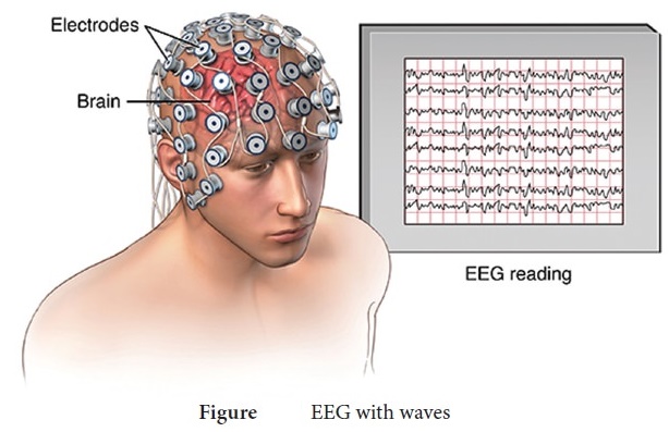
EEG with waves
14. Write the clinical significance of ultra sonogram
•
Ultrasound waves are used to image the foetus at different stages of pregnancy to
study the progress of the developing foetus.
•
They are used to hear foetal heart sound, blood flow, etc.
•
Used in echocardiography to diagnose the damages in heart.
•
Used for diagnosis of tumours, gall stones, kidney stones, obstructions in the genital
tracts.
15. Explain the principle involved in PET scan
Positron emission tomography Scanning (PET):
• PET is also computerized imaging technique unlike CT.
• Positron emission tomography (PET) is a nuclear medicine procedure
based on the measurement of positron emission from radiolabelled tracer molecules.
• These radiotracers allow biologic processes to be measured
and whole body images to be obtained which demonstrates sites of radiotracer accumulation.
• A PET image gives quantitative regional information on the
metabolic physiological processes.
• PET uses positron emitting radio isotopes (11C
13N 15O 18F) which are generated by the cyclotron.
• The most common radiotracer in use today is 18F-fluorodeoxyglucose
(18F-FDG) which is a radio labelled sugar (glucose) molecule.
• These atoms are then incorporated by chemical methods into
biological molecules like glucose, amino acids and ammonia.
• These positron emitting compounds are then injected in very
small amounts into or inhaled by experimental animals or human subjects.
• The three dimensional distribution of the labeled trace is
then probed by powerful PET cameras and the images are reconstructed by a computer.
• The quantitative interpretation of the image is done
by varying mathematical models.
• They deal with the process of uptake and metabolism of the radioisotope.
Related Topics