Chapter: Medical Microbiology: An Introduction to Infectious Diseases: Flagellates
Giardia lamblia - Parasitology
Giardia lamblia
PARASITOLOGY
G. lamblia was first described by Anton von Leeuwenhoek 300 years ago when he exam-ined his own diarrheal stool with one of the first primitive microscopes. It was not until the past several decades, however, that this cosmopolitan flagellate became widely re-garded in the United States as a pathogen. Of the six other flagellated protozoans known to parasitize the alimentary tract of humans, only one, Dientamoeba fragilis, has been credibly associated with disease. Definitive confirmation or refutation of its pathogenicity will, it is hoped, not require the passage of another three centuries.
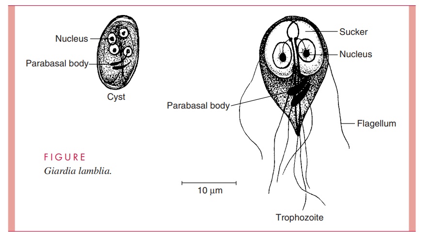
Unlike T. vaginalis, Giardia possesses both a trophozoite and a cyst form (Fig 54 – 2). It is a sting-ray – shaped trophozoite 9 to 21μm in length, 5 to 15μm in width, and 2 to 4μm in thickness. When viewed from the top, the organism’s two nuclei and central parabasal bodies give it the appearance of a face with two bespectacled eyes and a crooked mouth. Four pairs of flagella — anterior, lateral, ventral, and posterior — reinforce this image by suggesting the presence of hair and chin whiskers.
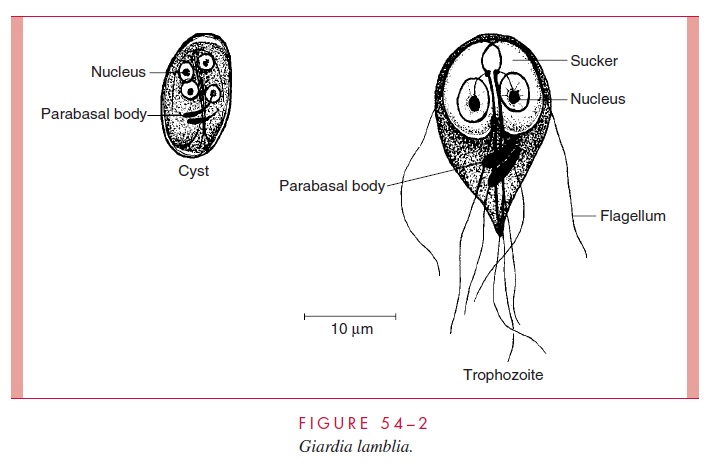
These distinctive parasites re-side in the duodenum and jejunum, where they thrive in the alkaline environment and ab-sorb nutrients from the intestinal tract. They move about the unstirred mucous layer at the base of the microvilli with a peculiar tumbling or “falling leaf ” motility or, with the aid of a large ventral sucker, attach themselves to the brush border of the intestinal epithelium. Unattached organisms may be carried by the fecal stream to the large intestine.
In the descending colon, if transit time allows, the flagella are retracted into cytoplas-mic sheaths and a smooth, clear cyst wall is secreted. These forms are oval and somewhat smaller than the trophozoites. With maturation, the internal structures divide, producing a quadrinucleate organism harboring two sucking discs, four parabasal bodies, and eight axonemes (see Fig 54 – 2). When fixed and stained, the cytoplasm pulls away from the cyst wall in a characteristic fashion. The mature cysts, which are the infective form of the parasite, may survive in cold water for more than 2 months and are resistant to concentra-tions of chlorine generally used in municipal water systems. They are transmitted from host to host by the fecal – oral route. In the duodenum of a new host, the cytoplasm di-vides to produce two binucleate trophozoites.
Organisms of the genus Giardia are among the most widely distributed of intestinal protozoa; they are found in fish, amphibians, reptiles, birds, and mammals. At first, it was assumed that Giardia strains found in different animals were host specific; on this basis, some 40 different species were described. As it is now recognized that some strains can infect multiple animal hosts, the practice of assigning species status by the host from which the parasite was recovered is considered invalid. Unfortunately, there is still no general agreement on an alternate method of speciation. Three morphologically distinct groups of Giardia have been described on the basis of their central parabasal body morphology.
Related Topics