Chapter: Medical Electronics : Electro-Physiology and Bio-Potential Recording
Electro Encephalo Gram (EEG)
ELECTRO ENCEPHALO
GRAM (EEG)
·
EEG is the recorded re presentation of bioelectric
potentials generated by the neuronal activity of the brain.
·
Basically, the brain is a gelatinous mass suspend
in the meanings, the cerebrospinal fluid, skull and scalp.
·
The brain is composed of three major subdivisions:
1. Cerebellum,
2. Brainstem
3. (Medulla, pons , midbrain, diencephalon) and
4. Cerebrum
The
cerebellum is mainly involved with skeletal muscle functions and maintenance
of balance. It coordinates smooth and directed movements.
·
The brain stemis the stalk of the brain and serves
as a relay station for all afferent (sensory) and efferent (motor) nerve fibers
between the spinal cord and higher brain canters. It also gives rise to ten of
the twelve cranial nerves, which supply the muscles and glands of the head and
major organs in the thoracic and abdominal cavities
·
Throughout the entire brainstem runs a core of
tissue called the reticular formation, which serves as a highly complex cluster
of neurons involved in integration of information from many afferent pathways
as well as from numerous other parts of the brain.
·
The cerebrum consists of the right and left
hemispheres. The outer part of the cerebral hemispheres, the cerebral cortex,
is a cellular shell 1.5 – 4 mm thick of grey matter.
·
The cerebral cortex is highly convoluted and is the
most complex integrating center of the nervous system. It brings together basic
sensory information into meaningful perceptual images and formulates ultimate
decisions for control over the motor systems of the body.
·
The cerebral cortex is comprised of two layers: the
pale cortex and the neocortex.
·
The pale cortex is located on the median surface
and the base of the brain and the neocortex is present on the superior and
lateral aspects of the cerebral hemispheres.
·
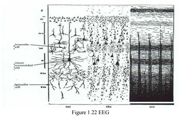
The neocortex is composed of six layers and its
cells can be categorized as pyramidal and non-pyramidal cells. There are
approximately 1010 neurons in the human cerebral cortex, about 75% of, which is
pyramidal.
· Pyramidal cells, named originally after their shape, have several characteristics. Their cell bodies are commonly triangular in shape, with the base down and the apex directed toward the cortical (superficial) surface.
·
The cell bodies vary in size, from axial dimensions
of 15 x 10 μm up to 120 x 90 μm. A typical pyramidal cell consists of a long
apical dendrite, about 2 mm long, that ascends from the apex of the cell body
and enters the overlaying layers and terminally branches within the outermost
layer of the neocortex.
· There is a dominant apical dendrites tree, looking like a forest of similarly oriented, densely packed units in the superficial layers of the neocortex, where extensive branching occurs.
·
There is also a basilar dendritic system that
extends out spherically from the cell body.
·
Pyramidal cells also have an axon that emerges from
the cell body and enters the sub cortical white matter.
· The axons of all pyramidal cells terminate in excitatory synapses. The initial segment of pyramidal cells is unmyelinated,as their recurrent branches
·
Axons of some pyramidal cells turn back toward the
cortical surface to end via their many dendritic branches on the dendrites of
other cells.
·
It has been shown by electrophysiological studies
that under normal circumstances, propagating action potentials in axons do not
contribute significantly to surface cortical recordings.
·
There reason being that action potentials travel in
large number of axons (running in many different directions relative to the
surface) in a temporally a synchronized way. Therefore, their net contribution
to the surface EEG is minimal and negligible.
·
It has been shown that the vertically oriented
pyramidal cells with their long apical dendrites running parallel to one
another are the major contributors to the electro genesis of the cortical field
potentials (EEG signal).
A highly schematic representation of a pyramidal cell and its role in the generation of surface EEG signal. Let’s consider a single pyramidal cell, and explain how potential changes in one part of the cell relative to other parts could generate the EEG signal.
·
Excitatory synaptic inputs to the branches in the
apical dendritic tree of the pyramidal cells cause depolarization of the
dendritic membrane.
·
This leads into generation of an excitatory
postsynaptic potential (EPSP)
·
As a result, a radially oriented dipole is set up
and sub threshold current flows in a closed path through the cytoplasmic core
of the dendrites and cell body of the cell, returning to the synaptic sites via
the conducting extracellular medium
·
The lines of current flow make the extracellular
medium close to the cell body act as a source with + polarity and the upper
part of the apical dendritic tree to act as a sink with – polarity.
·
This leads into recording a negative potential at
the cortical surface
In case
of inhibitory synaptic inputs to the branches in the apical dendritic tree, an
inhibitory postsynaptic potential (IPSP) is generated with a reversal in the
polarity of the current dipole, which leads into a generation of a positive
cortical recording.
·
Therefore, the influence of a particular dendritic
postsynaptic potential on the cortical recording depends on its net excitatory
or inhibitory effect and on its location relative to the measurement site.
The EEG
(electroencephalogram) signal is a recording of the electrical activity of the
brain. The EEG signal recorded at the cortex or the scalp is generated by the
polled activity of billions of cortical and sub cortical regions. The origin of
the EEG signal is based on the electrical activity of the pyramidal cells. The
EEG potentials primarily reflect the summated fluctuations of excitatory and
inhibitory postsynaptic potentials in the pyramidal cells of the upper layers
of the cerebral cortex. For reasons of geometry as well as because of extreme extracellular
attenuation, action potentials from firings of pyramidal cells contribute only
minimally or not all to the generation of the EEG signal.
·
All we need to contend ourselves with at this
stages that the EEG or brain waves are summation of neural depolarization sin
the brain due to the stimuli from the five senses as well as from thought
processes (indeed a very complex source). More on this in physiology in the
Nervous System topic.
·
EEG potentials have random-appearing waveforms with
peak-to-peak amplitudes ranging from less than 10 mV to over 100mV. Required
bandwidth is from below 1 Hz to over 100 Hz.
EEG is
recorded with 3 types of electrodes:
1. Scalp
2. Cortical
Electrocardiogram (recording from surface of cortex)
3. Depth
Electrodes recording from depth of brain (thin insulated needles of various
designs)
·
No matter where the recording is obtained from
(scalp, cortex or depth of the brain), the fluctuating potentials represent a
superposition of the volume conductor fields produced by a huge variety of
active neuronal current-generators.
·
On the surface of the brain (i.e.
Electrocardiogram), we can record voltages on the order of 10 mV! But, typical
EEG electrodes measure the electrical activity propagated through skull bone
and is attenuated from 1 to 100 μV.
·
EEG potentials vary as a function of position over
the surface of the skull, making it necessary to select sets of electrodes
grouped around Frontal, Parietal, Temporal and Occipital lobes.
The EEG Signal
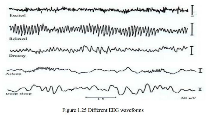
·
The character of the EEG signal is highly dependent
on the degree of the activity of the cerebral cortex, i.e. waves change
markedly between states of wakefulness and sleep.
·
Much of the time, EEGs are irregular and no general
pattern can be observed. Other times, distinct patterns emerge
·
The EEG waveform is divided into four wave groups:
1. The Alpha Waves (α) 8-13 Hz
2. The Beta
Waves (β) 14-30 Hz (The Gamma Waves (γ) 22-30 Hz or higher)
3. The Theta
Waves (θ) 4-7 Hz
4. The Delta
Waves (δ) <3.5 Hz
Note: During periods of mental
activity, the waves usually become asynchronous rather than synchronous, so the magnitude of summed potentials decreases in
spite of cortical activity.
·
In general there is a relationship between cerebral
activity and the frequency of the EEG rhythm
·
Frequency increases progressively with higher
degrees of activity
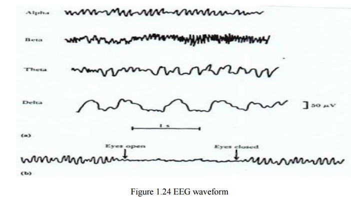
Examples:
· δ-Waves(<3.5 Hz) occur in surgical anesthesia and sleep
· θ-Waves(4-7 Hz) occur in emotional stress and frustration
·
α-Waves(8-13 Hz) occur during relaxed states
·
β-Waves(14-30 Hz)occur during intense mental
activity
The EEG
changes that occur as a human subject goes to sleep.
EEGs in Diagnosis
The
purpose of the clinical EEG is to help neurologists diagnose disease. The
pathological states most commonly diagnosed using EEG are:
·
Brain death (legal death)
·
Brain tumors
·
Epilepsy
·
Multiple Sclerosis
·
Sleep Disorder
·
Evoked responses (diseases of the audio, visual and
tactile senses)
·
Modern life sustaining equipment like respirators,
kidney dialyzers, ventilators, artificial heart pumps have changes the
definition of death
·
A sustained absence of EEG signal is a clinical
measure of brain death and can be used in deciding whether to transplant a
heart, liver, or lung or whether to shut down the life sustaining equipment
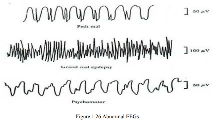
Some Representative Abnormal EEGS
Petit mal epilepsy– Minor for of seizure, clouding of
consciousness and loss of contact with the
environment
Grand mal epilepsy– Sudden loss of consciousness, falling
down, tonic contractions (stiffening of
muscles) followed by twitching and jerking movements of the limbs
Psychomotor seizures are
parietal seizures characterized by: semi-purposeful movements, changes in consciousness,
hallucinations and illusions.
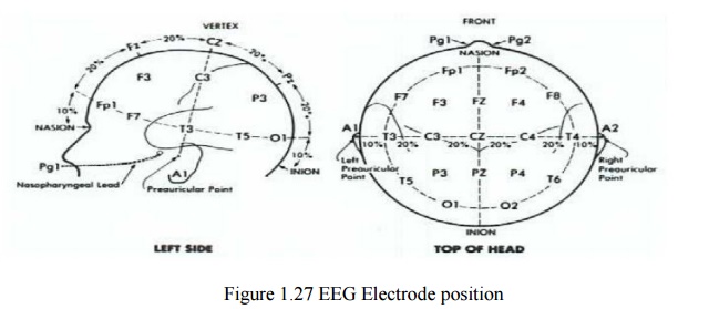
EEG Electrode Positions
·
In electroencephalography, the electrodes are
placed in an arrangement referred to as the 10-20 system
·
This is a placement scheme devised by the
International Federation of Societies of Electroencephalography
·
The electrodes are placed along a line drawn on the
skull from the root of the nose, the nasion, to the classification (bump on the
occipital lobe)
·
The first mark is placed 10% of the distance along
this line and others are arranged at 20% intervals
Electroencephalograph Signal Path
The EEG
signal path is comprised of: Scalp (biosignal source) EEG electrodes , Junction
box ,channel selector , differential amplifier, bank filters, display .
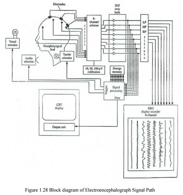
·
It shows the modern 8 channel EEG recorder. The
patient cable consists of 21 electrodes and is connected to the 8 channel
selector.
·
The electrodes are attached to the channel selector
in groups of 8 called a montage of electrodes.
·
The right ear electrode acts as reference electrode
for the right brain electrodes and left ear electrode act as reference
electrode for left brain electrodes.
·
The 50 Hz interference is reduced by employing
differential amplifiers as preamplifiers with more than 80 dB CMRR and by use
of 50 Hz notch filters.
·
The effect of notch filter on signal distortion is
not so much because important EEG signals have frequencies below 30 Hz.
·
The output voltage from the amplifier may either be
applied directly to the eight channel display through the filter bank or it may
be stored as data on a tape recorder or in a computer memory for further
processing.
Related Topics