Chapter: Essential Anesthesia From Science to Practice : Clinical management : Monitoring
Doppler and ultrasound - Anesthesia Clinical management

Doppler and ultrasound
The
Doppler principle has been applied to monitoring in anesthesia. We can place a
Doppler pencil probe over a vessel to identify blood flow or, with a broader
emitter/receiver head, place it over the chest to detect the blood flow in the right
When air appears in the blood flowing into the heart, it changes the reflective
characteristics of the blood, easily detected by the Doppler signal, which is
transformed into a swooshing noise.
While
ultrasound has been used for many years to spy on babies still in the womb and
to view the functioning heart through the chest wall, more recently the
equipment has been miniaturized into a finger-sized probe that views the heart
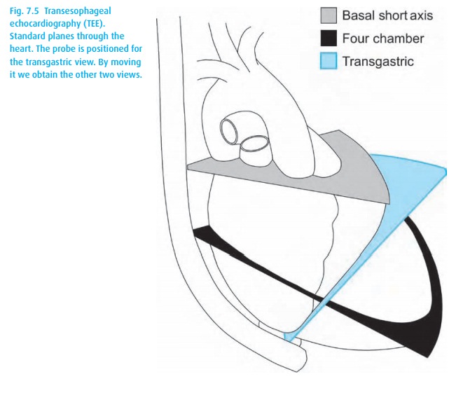
from behind, through the wall of the esophagus (Fig. 7.5).
This advance gives us a hands-free, relatively stable (and relatively
non-invasive) view of the heart that does not impinge on the operative field.
The technology continues to advance but currently allows views from multiple
angles and Doppler analysis of flow through the valves and even the coronaries
(for the experienced ultrasonographer). While invasive pressure monitoring can
give indirect insights into cardiac physiology, with TEE we can actually see the heart doing its work. We can
assess preload (how full is the ventricle?), contractility (how much are the
walls thickening?), and ischemia (are there sections of the ventricular walls
that lag behind?). During cardiac surgery, we can evaluate valve repairs and
ASD closures. TEE is also a great way to detect air emboli.
Related Topics