Chapter: Nervous System and Sensory Organs : Development and Structure of the Human Brain
Development of the Brain
Development of the Brain
The
closure of the neural groove into the neural tube begins at the level of the
upper cervical cord. From here, further closure runs in the oral direction up
to the rostral end of the brain (oral neuropore, later the terminal lamina) and
in the caudal direction up to the end of the spinal cord. Further developmental
events in the CNS proceed in the same directions. Thus, the brain’s divi-sions
do not mature simultaneously but at intervals (heterochronous maturation).
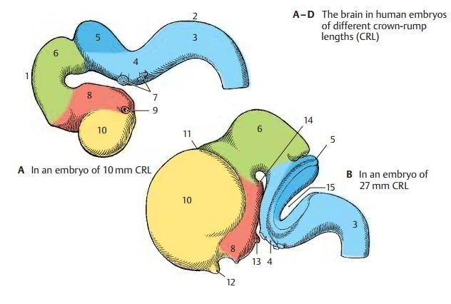
The
neural tube in the head region expands into several vesicles (p. 171, A). The
rostral vesicle is the
future forebrain, prosen- cephalon (yellow and red); the caudal
ves- icles are the future brain stem, encephalic trunk (blue). Two curvatures
of the neural tube appear at this time:
the cephalic flexure (A1) and the
cervical flexure (A2). Although the brain stem still shows a uni- form
structure at this early stage, the future divisions can already be identified:
medulla oblongata(elongatedcord) (A – D3), pons (bridge of Varolius)(A – D4), cerebellum (A – D5, dark blue), and
mesencephalon (mid- brain) (A – C6, green). The brain stem is
developmentally ahead of
the prosen- cephalon; during the
second month of human development, the telencephalon is still a thin-walled
vesicle (A), whereas neu- rons have already differentiated in the brain stem
(emergence of cranial nerves) (A7). The optic vesicle develops from the
diencephalon (AB8, red) (p. 343, A) and forms the optic cup (A9). Anterior to
it lies the telencephalic vesicle (telencephalon) (A – D10, yellow); ini-
tially, its anlage is unpaired (impar
telen-cephalon), but it soon expands on both sidesto form the two cerebral
hemispheres.
During
the third month, the prosen-cephalon enlarges (B). Telencephalon and diencephalon become separated by the telodiencephalic sulcus (B11). The anlage ofthe olfactory bulb (B – D12) has formed at
the hemispheric vesicle, and the pituitaryanlage (B13) (p. 201 B) and the
mamillary eminence (B14) have formed at the base of the diencephalon. A deep
transverse sulcus (B15) is formed between the cerebellar an- lage and the
medulla oblongata as a result of the pontine flexure; the underside of the
cerebellum comes to lie in apposition to the membrane-thin dorsal wall of the
medulla (p. 283, E).
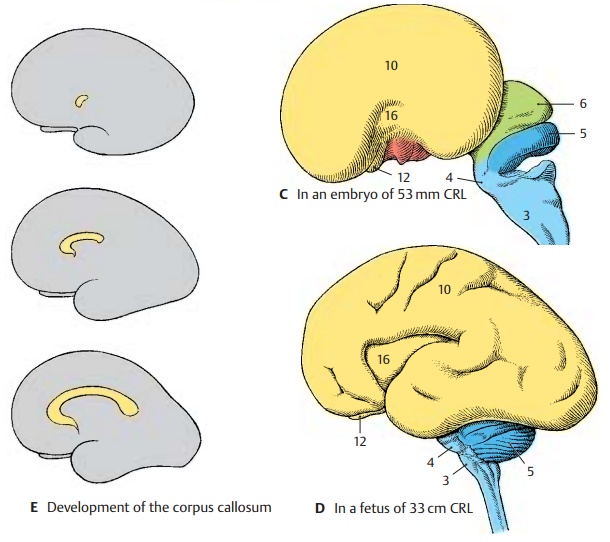
During the
fourth month, the
cerebral hemispheres begin to overgrow the other parts of the brain (C).
The telencephalon, which initially lagged behind all other brain divisions in
its development, now exhibits the most intense growth (p. 170, A). The center
of the lateral surface of each hemi- sphere lags behind in growth and later be-
comes overlain with parts. This is the insula (CD16). During the sixth month,
the insula still lies free (D). The first grooves and con- volutions appear on
the previously smooth surfaces of the hemispheres. The initially thin walls of
neural tube and brain vesicles have thickened during development. They contain
the neurons and nerve tracts that make up the brain substance proper. (For
development of cerebral hemispheres, see p. 208.)
Within
the anterior wall of the impar telen- cephalon, nerve fibers run from one hemi-
sphere to the other. The commissural sys- tems, which connect the two
hemispheres, develop in this segment of the thickened wall, or commissural
plate. The largest of them is the corpus callosum (E). The hemi- spheres grow
mainly in the caudal direc- tion; in parallel with their increase in size, the
corpus callosum also expands in the caudal direction during its development and
finally overlies the diencephalon.
Related Topics