Chapter: Human Nervous System and Sensory Organs : Cerebellum
Cerebellar Peduncles and Nuclei - Structure of Cerebellum

Cerebellar Peduncles and Nuclei
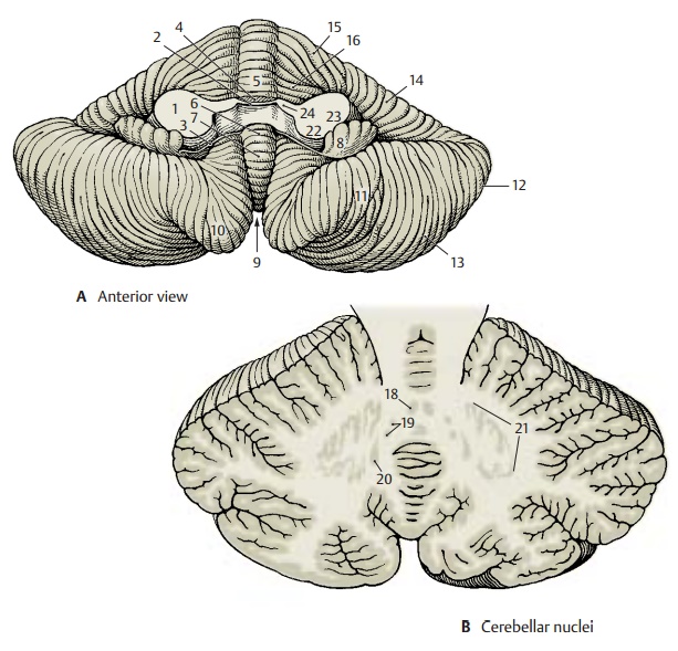
Anterior Surface (A)
On both sides, the cerebellum is connected to the brain stem by the cerebellar peduncles (A1). All afferent and efferent pathways pass through them. The anterior surface of the cerebellum becomes fully visible only after cutting through the peduncles and re-moving the pons and medulla oblongata. Between the cerebellar peduncles lies the roof of the fourth ventricle with the superiormedullary velum (A2) and the inferior medullary velum (A3). The anterior parts ofthe vermis are exposed, namely, the lingual (A4), the central lobule (A5), the nodulus(A6), the uvula (A7), and also the flocculus (A8). The vallecula of the cerebellum (A9) is surrounded on both sides by the tonsillae (A10).
The following parts are also visible: biven-tral lobule (A11), superior semilunar lobule (A12), inferior semilunar lobule (A13), simple lobule (A14), quadrangular lobule (A15), and wing of the central lobule (A16).

Nuclei (B)
The cross section shows cortex and nuclei of the cerebellum. The sulci are heavily branched, resulting in a leaflike configura-tion of the cross sectioned folia. The sagittal section thus shows a tree-like image, the arbor vitae (tree of life) (C17).
Deep in the white matter are the cerebellar nuclei. The fastigial nucleus (B18) lies close to the median line in the white matter of the vermis. It receives fibers from the cortex of the vermis, the vestibular nuclei, and the olive. It sends fibers to the vestibular nuclei and other nuclei of the medulla oblongata. The globose nucleus (B19), too, is thought to receive fibers from the cortex of the vermis and to send fibers to the nuclei of the medulla oblongata. Fibers of the cerebellar cortex from the region between vermis and hemisphere (intermediate part) are thought to terminate at the hilum of the dentate nu-cleus in the emboliform nucleus (B20). The fibers of the latter nucleus run through thesuperior cerebellar peduncle to the thalamus. The dentate nucleus (B21) appears as a heavily folded band with the medial part remaining open (hilum of dentate nu-cleus). The cortical fibers of the hemisphereterminate in the dentate nucleus, and fibers extend from here as superior cerebellar peduncle to the red nucleus and to the thalamus.

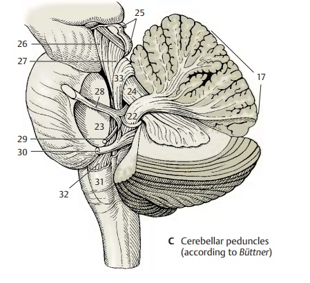
Cerebellar Peduncles (A, C)
The efferent and afferent pathways of the cerebellum run through three cerebellar peduncles:
· The inferior cerebellar peduncle (restiform body) (AC22), which ascends from the lower medulla oblongata; it contains the spinocerebellar tracts and the connec-tions to the vestibular nuclei
· The medial cerebellar peduncle (brachium pontis) (AC23) with the fiber masses from the pons, which originate from the pontine nuclei and represent the con-tinuation of the corticopontine tracts
· The superior cerebellar peduncle (brachium conjunctivum) (AC24), which contains the efferent fiber systems extending to the red nucleus and the thalamus
C25 Tectal plate.
C26 Medial lemniscus.
C27 Lateral lemniscus.
C28 Trigeminal nerve.
C29 Facial nerve.
C30 Vestibulocochlear nerve.
C32 Olive.
C32 Central tegmental tract.
C33 Anterior cerebellar tract.
Related Topics