Chapter: Medicine and surgery: Gastrointestinal system
Carcinoid tumours of the intestine - Gastrointestinal oncology
Carcinoid tumours of the intestine
Definition
Tumours originating from neuroendocrine or APUD (amine precursor uptake and decarboxylation) cells of the intestine.
Aetiology/pathophysiology
Carcinoid tumours most commonly occur in the appendix, terminal ileum and rectum. Small bowel, colonic and stomach lesions grow slowly and metastasise to the liver. Rectal and appendix carcinoids almost never metastasise.
Clinical features
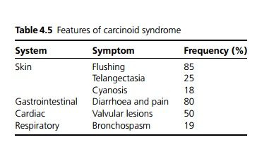
Most lesions are asymptomatic although appendix carcinoids may present with acute appendicitis. Carcinoid syndrome occurs in 5% with liver metastases, the features of which (see Table 4.5) depend on the hormones produced.

Macroscopy/microscopy
Carcinoid tumours appear as a yellow – white mural nodule composed of cords and nests of cells with secretory granules.
Investigations
Plasma chromogranin A is raised in carcinoid tumours. Urinary 5-hydroxyindoleacetic acid (a 5 HT metabolite) is useful for carcinoid tumours of the midgut. Staging investigations include octreotide scanning, liver ultra-sound, spiral CT scanning, MRI scanning and endoscopic ultrasound.
Management
Resection of the primary tumour (e.g. by appendicectomy) is performed where possible. Octreotide (somatostatin analogue) relieves diarrhoea and flushing and may reduce tumour growth. Other treatments include chemotherapy, embolisation and thermal ablation.
Related Topics