Chapter: Maternal and Child Health Nursing : Anatomy and physiology of female reproductive
Bony Pelvis
The Bony Pelvis
The bony
pelvis form the bony canal through which the fetus must pass during the normal
birth process. If the canal is of the normal shape, and size, the baby of the
normal size will negotiate it without difficulties,because pelvis vary in size
and shape it is important that the midwife recognizes the normal pelvis so as
to be able to detect deviation from the normal. One of the ways of estimating
the progress of labor is by assessing the relationship of the fetus to certain
pelvic landmarks.
Functions
·
It connects the spine to the lower limbs
·
It protects the female reproductive organs,
bladder, the urethra ,colon, rectum and anal canal
·
It allows movement of the body especially walking
and running
·
It permits sitting
and kneeling
·
It forms a bony passage for the fetus during labor
·
It transmits the weight of the trunk to the legs
and holds the two femurs
·
Protects the pelvic organs and to a lesser extent
the abdominal contents
·
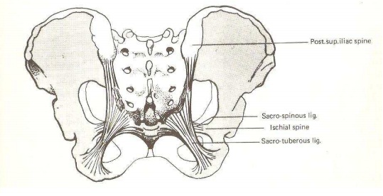
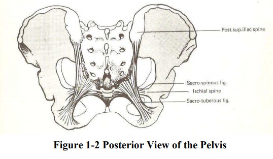
The Sacrum transmits cauda equina and distributes nerves to various parts of the pelvis
Pelvic bones: Thereare four
bones in the pelvis
·
Two innominate bones (hip bones),
·
One sacrum and
·
One coccyx
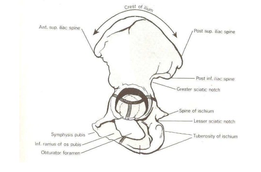
InnominateBones:
each innominate bone is composed of three parts - the ilium, ischium and the
pubis
·
Ilium – large flared out part
·
Ischium – the thick lower part
·
Pubic bone – forms the anterior part
Sacrum is
a wedge shaped bone consisting of five fused vertebrae. Upper border of the
first sacral vertebral juts forward, known as SacralPromontory which is the most importantlandmark in the female
pelvis. Anterior surface is concave, referred to as Hollow ofthe Sacrum. Lateral sacrum extends into a wing or ala. Posterior surface is
roughened toreceive attachment of muscles. Two pairs of holes, or foramina,
pierce the sacrum through which, nerve from the cauda equina emerge to supply
pelvic organs.
Coccyx is
a vestigial tail. It consists of four fused vertebrae forming a small
triangular bone. The coccyx bends backwards at this joint during parturition to
increase the anterior posterior diameter of the pelvic outlet.
Pelvic Joints - there are four pelvic joints
·
One symphysis pubis – formed at the joint of two
pubic bones, united by a pad of cartilage known as the symphysis pubis
·
Two (right and left) sacroiliac joints – is the
strongest joint in the body articulates sacrum to ilium. Normally there are
little or no movements in these joints, but during pregnancy especially towards
the end there is a certain degree of movement due to the relaxation of the
ligaments of the joints. This may give rise to difficulties in walking and
backache, especially the multiparous women. There is little widening during
labour , commonly referred to as “give” of the pelvis.
·
One sacrococcygeal joint – join the base of the
coccyx to the tip of the sacrum
Pelvic Ligaments: ligaments bind the joints
·
Inter pubic ligaments at the symphysis pubis
·
Sacroiliac ligaments.
·
Sacrotuberous ligament
·
Sacrococcygeal ligaments.
·
Inguinal ligament
Division of the Pelvis
The
pelvis is divided into two parts, the true and the false pelvis. The false is
the part above the brim. It has little importance in obstetrics
The true pelvis is the bony canal through whichfetus
must pass during birth. It consists of brim, cavity and outlet. Brim is round
except where sacral promontory projects into it. Commencing posteriorly the
pelvic brim includes the following important landmarks.
·
Sacral promontory
·
Sacral ala or wing
·
Sacroiliac joints
·
Iliopectineal line
·
Iliopectineal eminence
·
Superior ramus of the pubic bone
·
Upper inner border of the body of the pubic bone
·
Upper inner border of the symphysis pubis
Diameters of the pelvis
Diameters
of the brim
·
Antero-posterior diameter – from sacral promontory
to upper most border of symphysis pubis 12cm. A measurement to the posterior
border of the upper surface to a point 1.25cm lower is called the obstetrical
conjugate, 11cm. It is the available space for the passage of the fetus hence
it is called the true conjugate
·
Diagonal conjugate is anteroposterior diameter from
the lower border of the symphysis pubis to the centre of the sacral promontory
measured vaginally for pelvic assessment 12-13 cm.
·
Oblique diameter – from sacroiliac joint to the
iliopectineal eminence on the opposite side (right and left). It measures 12cm
·
Transverse diameter – it is between the points
furthest apart on iliopectineal lines and measures 13cm. The fetal head
commonly enters in transverse diameter of the pelvic brim
·
Sacrocotyloid diameter – from sacral promontory to
the iliopectineal eminence on each side, measures 9 – 9.5 cm
·
The pelvic cavity extends from the pelvic brim
above to the outlet below. Anterior wall is formed by pubic bones and symphysis
pubis - depth is 4cm. The cavity is circular in shape and is considered to be
12cm all round.
Diameter
of the outlet:
There are
two Pelvic outlets: described as Anatomical
Outlet and Obstetrical Outlet.
Theanteroposterior diameter of outlet – from the lower border of the symphysis
pubis to the sacrococcygeal joints 13cm
The oblique diameter of outlet - from the
oburatorforamen to the sacrospinous ligament 12cm
The transverse diameter of outlet - is
takenbetween two ischial spines 10 -11 cm which is the narrowest diameter of
the pelvis
Pelvic inclination there is
difference in theinclination of the pelvis when the woman is standing, sitting
and recumbent position. The inclination of the outlet is 11O, cavity
30O, brim 60O, almost 90O in Negro woman
Pelvic planes these are imaginary flat
surfacesat the brim, cavity and outlet of the pelvic canal. The fetus will
enter at right angle to the plane according to the inclination.
Axis of the pelvic canal a line
drawn exactly halfway between anterior wall and posterior wall of the pelvic
canal to the plan of the outlet, cavity and the brim the curve it makes is
known as the curve of Carus, the path which the fetus takes as it travels
trough the birth canal.
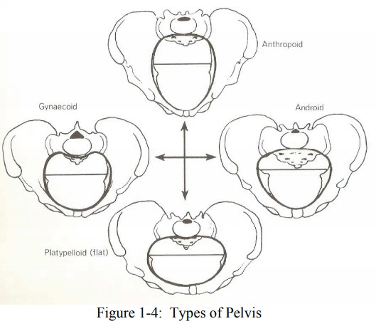
Types of Pelvis
·
Gynaecoid pelvis – ideal pelvis for child bearing
·
Android pelvis – resembles a male pelvis
·
Anthropoid pelvis – has long oval brim in which
anteroposterior diameter is longer than transverse diameters. Labor does not
present any difficulties but favors occipitoanterior or occipitoposterior
positions
·
Platypelloid pelvis - flat with kidney shaped brim
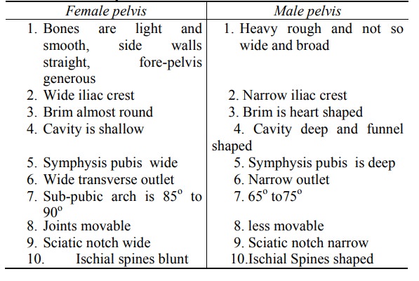
Comparison of Male and Female Pelvis
Related Topics