Chapter: Medical Immunology: Organ-Specific Autoimmune Diseases
Autoimmune Diseases of the Blood Cells
AUTOIMMUNE DISEASES OF THE BLOOD CELLS
Virtually all types of blood cells can be affected by autoantibodies.
A. Autoimmune Neutropenia
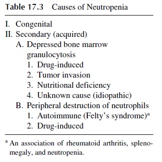
A reduction of the total number of neutrophils is the most frequent cause of infection due to defective phagocytosis. Although there are congenital forms of neutropenia of variable severity, most frequently neutropenia is secondary to a variety of causes (see Table 17.3). Autoimmune neutropenia can be seen in patients with rheumatoid arthritis, usually in as-sociation with splenomegaly (Felty’s syndrome).

B. Idiopathic Thrombocytopenic Purpura
Idiopathic thrombocytopenic purpura (ITP) is an autoimmune disease associated with low platelet counts (thrombocytopenia). The low platelet counts result from a shortened platelet half-life, usually caused by antiplatelet antibodies that cannot be compensated by increased release of platelets from the bone marrow.
Antiplatelet autoantibodies have been detected in 60–70% of patients with the “im-munoinjury” technique that relies on the release of platelet factors, such as serotonin, fol-lowing exposure to sera containing such antibodies. Competitive binding assays and antiglobulin assays can also be used to demonstrate antiplatelet antibodies.
ITP can present as an acute or as a chronic form. Acute ITP is due to the formation of immune complexes containing viral antigens that become adsorbed to the platelets or to the production of antiviral antibodies that cross-react with platelets. Platelet destruction can be due to irreversible aggregation caused by immune complexes, or when antiplatelet antibod-ies are involved, or to complement-induced lysis or phagocytosis. Chronic ITP is caused by autoantibodies that react with platelets and lead to their destruction by phagocytosis.
Clinically, ITP is characterized by easy and exaggerated bleeding secondary to thrombocytopenia. Patients with ITP usually present with subcutaneous and mucosal bleeding. The subcutaneous bleeding is clinically described as petechiae or ecchymosis, ap-pearing without obvious cause. When the bleeding is profuse, it will lead to the appearance of areas of purple discoloration, from which the designation of “purpura” derives. Epistaxis and gingival, genitourinary, and gastrointestinal tract bleeding are the common manifesta-tions of mucosal bleeding.
The acute forms of ITP are seen mainly in children, often in the phase of recovery af-ter a viral exanthem or an upper respiratory infection and is usually self-limited. In contrast, chronic ITP is an adult disease, often associated with other autoimmune diseases. The di-agnosis is based on the platelet count, which in the acute form is usually less than 20,000/mm3, whereas in the chronic form the platelet counts range between 30,000 and 100,000/mm3. The white cell count is usually normal. The bone marrow is also usually nor-mal, but in some cases an increase in megakaryocytes may be seen, representing an attempt to compensate for the excessive destruction in the peripheral blood. The spleen may be en-larged due to platelet sequestration by phagocytic cells.
The treatment of ITP can be surgical or medical. Splenectomy is usually reserved for patients in whom the spleen is the major site for platelet sequestration and destruction. The removal of the spleen is often associated with prolonged platelet survival.
Glucocorticoids have been used in cases in which splenectomy is not indicated or has not been beneficial. However, the efficiency of glucocorticoids in severe cases of ITP is questionable.
In the last decade, administration of intravenous gammaglobulin has become the therapy of choice. It is associated with a prolongation of platelet survival and improvement in platelet counts. Several mechanisms have been proposed to explain this effect of intra-venous gammaglobulin in ITP:
1. Competition with immune complexes for the binding to platelets (immune complexes would cause irreversible aggregation or complement-mediated cytolysis and intravenous gammaglobulin would not).
2. Blocking of Fc receptors in phagocytic cells, which would inhibit the ingestion and destruction of antibody-coated platelets (for which there is experimental documentation). This mechanism is most likely to explain the rapid increase in platelet counts seen after therapy with intravenous immunoglobulin is initiated.
3. The long-term, beneficial effects of intravenous gammaglobulin administration are apparently a consequence of the downregulation of autoreactive B cells caused by co-ligation of mlg (by anti-idiotypic antibodies reactive with the mlg of autoreactive B cells present in the IVIg) and FcIIÎłR (by the Fc region of IVIg). This co-ligation is believed to even be able to induce B-cell apoptosis. The end result is a decrease of the titers of antiplatelet antibodies.
Related Topics