Chapter: Human Neuroanatomy(Fundamental and Clinical): Basic Neuronal Arrangements
Afferent Neurons
Afferent Neurons
We have seen that afferent nerve fibres can be divided into four categories viz., general somatic afferent, special somatic afferent, general visceral afferent and special visceral afferent. The basic arrangement of the neurons that give origin to all four categories of afferent fibres is similar, and the description that follows applies to all of them.
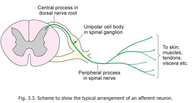
The cell bodies of neurons that give rise to efferent fibres of peripheral nerves are located within the brain and spinal cord. In contrast, the cell bodies of neurons that give rise to afferent fibres are located outside the CNS. In the case of spinal nerves the cell bodies lie in the spinal ganglia; and in the case of the cranial nerves they lie in sensory ganglia (e.g., the trigeminal ganglion) associated with these nerves. We may illustrate the arrangement with reference to a spinal nerve (Fig. 3.3). The cells of the dorsal nerve root ganglion are of the unipolar variety described. Each cell gives off a single process that divides into a peripheral process and a central process. The peripheral process extends into the spinal nerve and courses through its branches to reach the tissue or organ supplied. It may branch repeatedly during its course. We have already seen that these peripheral processes are functionally dendrites as they convey impulses towards the cell body, but they are indistinguishable in structure from axons. These processes constitute the sensory fibres of peripheral
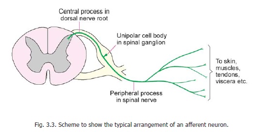
nerves. The sensory impulses brought by these processes from various organs of the body are conveyed to the spinal cord by the central processes (representing axons). Within the spinal cord the central processes usually run a short course and terminate by synapsing with cells in the posterior grey column. Some of the central processes are, however, long. They enter the posterior funiculus and run upwards to the medulla as ascending tracts.
The sensory ganglia of the fifth, seventh, ninth and tenth cranial nerves are made up of cells similar to those of the spinal ganglia. Their central processes end by synapsing with cells in the sensory nuclei of these nerves. The sensory ganglia of the eighth nerve (i.e., the cochlear and vestibular ganglia) are peculiar in that their neurons are bipolar, the two processes corresponding to the central and peripheral processes of unipolar neurons.
Related Topics