Chapter: Medicine and surgery: Nervous system
Abnormalities of the optic disc - Disorders of cranial nerves
Abnormalities of the optic disc
Definition
The optic disc is where the retinal fibres meet to form the optic nerve. Diseases affecting the optic nerve may cause the disc to look abnormal:
i. Swollen, i.e. less cupped – papilloedema, optic neuritis (sometimes called papillitis)
ii. Pale due to loss of axons and vascularity – optic atrophy
Papilloedema
This term should be reserved to describe swelling of the optic disc due to raised intracranial pressure (or pressure behind the eye). The increased pressure causes axonal transport to become abnormal, causing swelling of the nerves. Papilloedema is usually (not always) bilateral, there is loss of venous pulsation, visual acuity is preserved (but with constriction of visual fields and an enlarged blind spot).
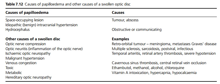
The term is often used to cover all causes of a swollen disc, but this is the differential diagnosis of papilloedema (see Table 7.12).

Optic atrophy
Optic atrophy may follow any damage to the optic nerve, particularly after ischaemia, optic neuritis and optic nerve compression. It may also be hereditary.
Clinical features
The degree of visual loss depends on the underlying cause. Optic neuritis and ischaemic neuropathy typically cause early visual loss.
Management
Directed at the underlying cause.
Related Topics