Chapter: Medical Physiology: The Eye: III. Central Neurophysiology of Vision
ÔÇťFusionÔÇŁ of the Visual Images from the Two Eyes
ÔÇťFusionÔÇŁ of the Visual Images from the Two Eyes
To make the visual perceptions more meaningful, the visual images in the two eyes normally fuse with each other on ÔÇťcorresponding pointsÔÇŁ of the two retinas. The visual cortex plays an important role in fusion. It was pointed out earlier that correspon-ding points of the two retinas transmit visual signals to different neuronal layers of the lateral geniculate body, and these signals in turn are relayed to parallel neurons in the visual cortex. Interactions occur between these cortical neurons to causeinterferenceexcitation in specific neurons when the two visualimages are not ÔÇťin registerÔÇŁÔÇöthat is, are not precisely ÔÇťfused.ÔÇŁ This excitation presumably provides the signal that is transmitted to the oculomotor apparatus to cause convergence or divergence or rotation of the eyes so that fusion can be re-established. Once the cor-responding points of the two retinas are in register, excitation of the specific ÔÇťinterferenceÔÇŁ neurons in the visual cortex disappears.
Neural Mechanism of Stereopsis for Judging Distances of Visual Objects It is pointed out that because the two eyes are more than 2 inches apart, the images on the two retinas are not exactly the same. That is, the right eye sees a little more of the right-hand side of the object, and the left eye a little more of the left-hand side, and the closer the object, the greater the dispar-ity. Therefore, even when the two eyes are fused with each other, it is still impossible for all corresponding points in the two visual images to be exactly in regis-ter at the same time. Furthermore, the nearer the object is to the eyes, the less the degree of register. This degree of nonregister provides the neural mechanism for stereopsis, an important mechanism for judging the distances of visual objects up to about 200 feet (60 meters).
The neuronal cellular mechanism for stereopsis is based on the fact that some of the fiber pathways from the retinas to the visual cortex stray 1 to 2 degrees on each side of the central pathway. Therefore, some optic pathways from the two eyes are exactly in register for objects 2 meters away; still another set of pathways is in register for objects 25 meters away. Thus, the dis-tance is determined by which set or sets of pathways are excited by nonregister or register. This phenome-non is called depth perception, which is another name for stereopsis.
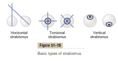
Strabismus
Strabismus, also called squint or cross-eye, means lack of fusion of the eyes in one or more of the visual coor-dinates: horizontal, vertical, or rotational. The basic types of strabismus are shown in Figure 51ÔÇô10: (1) hor-izontal strabismus, (2) torsional strabismus, and (3) ver-tical strabismus. Combinations of two or even all threeof the different types of strabismus often occur.
Strabismus is often caused by abnormal ÔÇťsetÔÇŁ of the fusion mechanism of the visual system. That is, in a young childÔÇÖs early efforts to fixate the two eyes on the same object, one of the eyes fixates satisfactorily while the other fails do so, or they both fixate satisfactorily but never simultaneously. Soon the patterns of conjugate movements of the eyes become abnormally ÔÇťsetÔÇŁ in the neuronal control pathways themselves, so that the eyes never fuse.
Suppression of the Visual Image from a Repressed Eye. In a fewpatients with strabismus, the eyes alternate in fixing on the object of attention. In other patients, one eye alone is used all the time, and the other eye becomes repressed and is never used for precise vision. The visual acuity of the repressed eye develops only slightly, sometimes remaining 20/400 or less. If the dominant eye then becomes blinded, vision in the repressed eye can develop only to a slight extent in adults but far more in young children. This demonstrates that visual acuity is highly dependent on proper development of central nervous system synaptic connections from the eyes. In fact, even anatomically, the numbers of neuronal con-nections diminish in the visual cortex areas that would normally receive signals from the repressed eye.
Related Topics