Chapter: Essentials of Anatomy and Physiology: Urinary System and Fluid Balance
Urine Movement
URINE MOVEMENT
Anatomy and histology of the Ureters, Urinary Bladder, and Urethra
The ureters are small tubes that carry urine from the renal pelvis of the kidney to the posterior inferior portion of the urinary blad-der (figure 18.18). The urinary bladder is a hollow, muscular container that lies in the pelvic cavity just posterior to the pubic symphysis. It stores urine; thus, its size depends on the quantity of urine present. The urinary bladder can hold from a few mil-liliters (mL) to a maximum of about 1000 mL of urine. When the urinary bladder reaches a volume of a few hundred mL, its wall is stretched enough to activate a reflex that causes the smooth muscle of the urinary bladder to contract, and most of the urine flows out of the urinary bladder through the urethra.
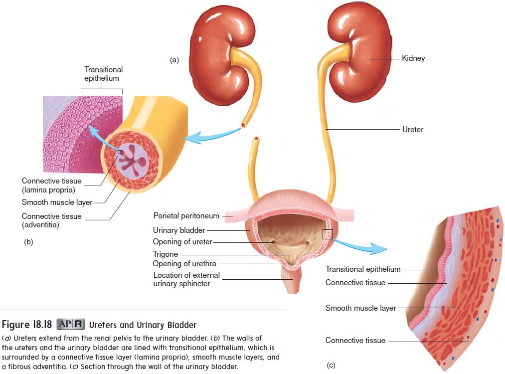
The urethra is the tube that carries urine from the urinary bladder to the outside of the body. The triangle-shaped portion of the urinary bladder located between the opening of the ureters and the opening of the urethra is called the trigone (trı̄′ gōn; triangle).
The ureters and the urinary bladder are lined with transitional epithelium, which is specialized to stretch . As the volume of the urinary bladder increases, the epithelial cells change in shape from columnar to flat, and the number of epithelial cell layers decreases. As the volume of the urinary bladder decreases, transitional epithelial cells assume their columnar shape and form a greater number of cell layers.
The walls of the ureter and urinary bladder are composed of layers of smooth muscle and connective tissue. Regular waves of smooth muscle contraction in the ureters produce the force that causes urine to flow from the kidneys to the urinary bladder. Contractions of smooth muscle in the urinary bladder force urine to flow from the bladder through the urethra.
At the junction of the urinary bladder and the urethra, the smooth muscle of the bladder wall forms the internal urinarysphincter in males. (There is no functional internal urinarysphincter in females.) In males, the internal urinary sphincter contracts to keep semen from entering the urinary bladder during sexual intercourse . In both males and females, the external urinary sphincter is formed of skeletal muscle that sur-rounds the urethra as the urethra extends through the pelvic floor. The external urinary sphincter is under voluntary control, allowing a person to start or stop the flow of urine through the urethra.
In males, the urethra extends to the end of the penis, where it opens to the outside. The female urethra is much shorter (approxi-mately 4 cm) than the male urethra (approximately 20 cm) and opens into the vestibule anterior to the vaginal opening.
Micturition Relex
The micturition (mik-choo-rish′ ŭn) reflex is activated by stretch of the urinary bladder wall. As the urinary bladder fills with urine, pressure increases, stimulating stretch receptors in the wall of the urinary bladder. Action potentials are conducted from the urinary bladder to the spinal cord through the pelvic nerves. Integration of the reflex occurs in the spinal cord, and action potentials are conducted along parasympathetic nerve fibers to the urinary blad-der. Parasympathetic action potentials cause the urinary bladder to contract.(figure 18.19)
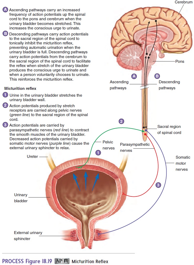
The external urinary sphincter is normally contracted as a result of stimulation from the somatic motor nervous system. Because of the micturition reflex, action potentials conducted along somatic motor nerves to the external urinary sphincter decrease, which causes the sphincter to relax. The micturition reflex is an automatic reflex, but it can be inhibited or stimulated by higher centers in the brain. The higher brain centers prevent micturition by sending action potentials through the spinal cord to decrease the intensity of the autonomic reflex that stimulates urinary bladder contractions and to stimulate nerve fibers that keep the external urinary sphincter contracted. The ability to voluntarily inhibit micturition develops at the age of 2-3 years.
When a person feels the urge to urinate, the higher brain centers alter action potentials sent to the spinal cord to facilitate the mic-turition reflex and relax the external urinary sphincter. Awareness of the need to urinate occurs because stretch of the urinary bladder stimulates sensory nerve fibers that increase action potentials car-ried to the brain by ascending tracts in the spinal cord. Irritation of the urinary bladder or the urethra by a bacterial infection or some other condition can also initiate the urge to urinate, even though the urinary bladder is nearly empty.
Related Topics