Chapter: Human Neuroanatomy(Fundamental and Clinical): The Olfactory and Limbic Regions
The Limbic Region
The Limbic Region
The term limbic system has been applied, in the past, to certain regions of the brain that are believed to play an important role in the control of visceral activity. Many of these areas have been considered as having a predominantly olfactory function; but it is now realised that this is not so. The areas of cerebral cortex included in the region are often referred to as the limbic lobe.
Some of the functions attributed to the so called limbic system are as follows.
a) Integration of olfactory, visceral, and somatic impulses reaching the brain.
b) Control of activities necessary for survival of the animal, including the procuring of food and eating behaviour.
c) Control of activities necessary for survival of the species including sex behaviour.
d) Emotional behaviour.
e) Retention of recent memory.
These can be interfered with in lesions of the region.
However, at present concepts regarding the unity of the so called limbic system are being seriously questioned, and the concept of these structures forming a “system” may well be discarded in future. It is for this reason that the heading for this section has been changed from the limbic system (in the previous edition) to the limbic region.
The areas to be included under the heading limbic system are not completely agreed upon. The following are generally included.
Areas forming the limbic cortex
One of the problems in identifying the areas to be included under this heading arises from the fact that much information has been derived from experiments on animals. The areas included, in the brains of higher primates, include the following.
a) Hippocampus (Ammon’s horn), and dentate gyrus.
b) Entorhinal cortex.
c) Gyrus cinguli (some parts of which are excluded), and paraterminal gyrus
d) Part of the parahippocampal gyrus.
e) The indusium griseum may be regarded as a vestigial part of the limbic cortex.
f) The amygdaloid nuclei are included under the limbic cortex. (This may appear strange as these nuclei are not part of the cerebral cortex. However, it has been found that the connections of these nuclei are similar to those of the cortex).
2. Bordering the limbic cortex there areperilimbic areas. These include those parts of the gyruscinguli and the parahippocampal gyrus that are not included in the limbic cortex. The retrosplenial gyrus (or gyrus fasciolaris) is also included.
3. Some authors include some of the olfactory areas in the region of the anterior perforated substance,and the septal region.
Fibre bundles related to the limbic region
These include the following
1. Olfactory nerves, tract and striae.
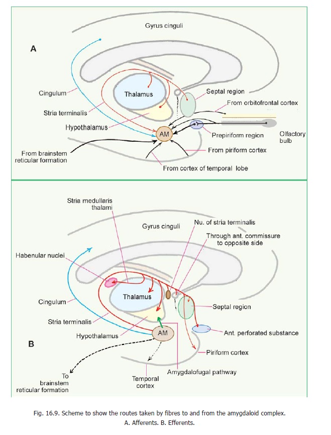
2. Fornix, stria terminalis, stria medullaris thalami, diagonal band, and anterior commissure.Some of these regions are considered in detail below. It is relevant to know that some regions not included in the limbic region, are nevertheless closely related to it functionally. These include the hypothalamus, and the reticular formation of the midbrain. The medial part of the thalamus, and the frontal cortex are also closely related functionally to the limbic system. Several of the areas included in the limbic region have been considered along with the olfactory pathway.
Amygdaloid Nuclear Complex
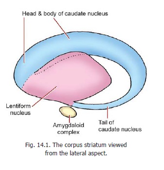
This region is also called the amygdaloid body or amygdala. Some features of this region have been considered. It is situated near the temporal pole of the cerebral hemisphere in close relation to the anterior end of the inferior horn of the lateral ventricle. Superiorly, the complex is related to the anterior part of the lentiform nucleus. Inferiorly, the complex is related to the gyrus semilunaris, the gyrus ambiens and the uncinate gyrus. It lies just below the anterior end of the tail of the caudate nucleus. The lower end of the stria terminalis lies in relation to the amygdaloid complex (Figs. 14.1 and 16.9).
Advanced
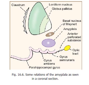
In the region between the amygdaloid complex and the lentiform nucleus there is a region of substriatal grey matter within which there is a collection of cholinergic neurons. These neurons form the basal nucleus of Meynert. Some relationships of the amygdaloid complex are shown in Fig. 16.6.
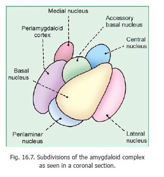
The amygdaloid complex is divided into a number of nuclei that have a complex terminology. A simplified version is shown in Fig. 16.7. Identify the lateral, medial, basaland central nuclei, and the periamygdaloid cortex. The basal and lateral nuclei are collectively referred to as the basolateralnucleus. Cells of the central and medial nuclei spread backwards along the stria terminalis and, alongwith this extension, they are referred to as the extended amygdala.
Related Topics