Chapter: Human Neuroanatomy(Fundamental and Clinical): Gross Anatomy of the Cerebellum
Subdivisions of the Cerebellum - Gross Anatomy of the Cerebellum
Gross Anatomy of the Cerebellum
The cerebellum (or small brain) lies in the posterior cranial fossa. In the adult the weight of the cerebellum is about 150 g. This is about ten per cent of the weight of the cerebral hemispheres. Like the cerebrum, the cerebellum has a superficial layer of grey matter, the cerebellar cortex. Because of the presence of numerous fissures, the cerebellar cortex is much more extensive than the size of this part of the brain would suggest. It has been estimated that the surface area of the cerebellar cortex is about fifty per cent of the area of the cerebral cortex.
The cerebellum lies behind the pons and the medulla. It is separated from the cerebrum by a fold of dura mater called the tentorium cerebelli. Anteriorly, the fourth ventricle intervenes between the cerebellum (behind), and the pons and medulla (in front). Part of the cavity of the ventricle extends into the cerebellum as a transverse cleft. This cleft is bounded cranially by the superior (or anterior) medullary velum, a lamina of white matter (Fig. 20.13).
Subdivisions of the Cerebellum
The cerebellum consists of a part lying near the midline called the vermis, and of two lateral hemispheres. It has two surfaces,superior and inferior. On the superior aspect, there is no lineof distinction between vermis and hemispheres. On the inferior aspect, the two hemispheres are separated by a deep depression called the vallecula. The vermis lies in the depth of this depression. On each side the vermis is separated from the corresponding cerebellar hemisphere by a paramediansulcus. Anteriorly and posteriorly the hemispheres extend beyond the vermis and are separated byanterior and posterior cerebellar notches. (The falx cerebelli lies in the posterior notch).
The surface of the cerebellum is marked by a series of fissures that run more or less parallel to one another. The fissures subdivide the surface of the cerebellum into narrow leaf like bands or folia. The long axis of the majority of folia is more or less transverse. Sections of the cerebellum cut at right angles to this axis have a characteristic tree-like appearance to which the termarbor-vitae (tree of life) is applied.
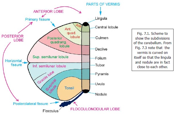
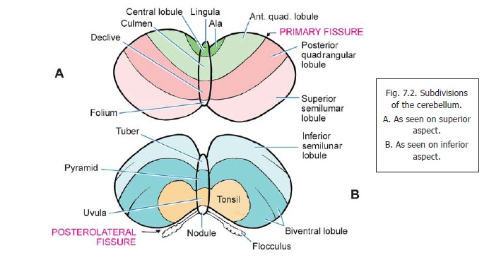
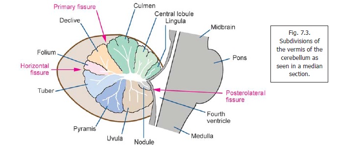
Some of the fissures on the surface of the cerebellum are deeper than others. They divide the cerebellum into lobes within which smaller lobules may be recognised. To show the various subdivisions of the cerebellum in a single illustration it is usual to represent the organ as if it has been ‘opened out’ so that the superior and inferior aspects can both be seen. Such an illustration is shown in Fig. 7.1. This should be compared with Figs. 7.2 A and B which are more realistic drawings of the superior and inferior surfaces, and with Fig. 7.3 which is a midline section showing the subdivisions of the vermis.
The deepest fissures in the cerebellum are:
i. the primary fissure (fissura prima) running transversely across the superior surface, and
ii. theposterolateral fissure seen on the inferior aspect.
These fissures divide the cerebellum into three lobes. The part anterior to the primary fissure is the anterior lobe. The part between the two fissures is the posterior lobe (sometimes called the middle lobe). The remaining part is the flocculonodular lobe.The anterior and posterior lobestogether form the corpus cerebelli.
The vermis is so called because it resembles a worm. Proceeding from above downwards in Fig. 7.1 it is seen to consist of thelingula, central lobule and culmen (in the anterior lobe); the declive,folium (or folium vermis), tuber (or tuber vermis), pyramis (or pyramid) and uvula (in themiddle lobe); and the nodule (in the flocculonodular lobe).
(The declive is sometimes referred to as “simple” as it is part of the simple lobule: see next para). With the exception of the lingula each subdivision of the vermis is related laterally to a part of the hemisphere. In the anterior lobe, we have the ala lateral to the central lobule; and the anteriorquadrangular lobule lateral to the culmen. (A very small part lateral to the lingula is called thewing of the lingula). In the middle lobe, we have the posterior quadrangular lobule lateral to the declive; the superior semilunar lobulelateral to the folium; the inferior semilunar lobule and the gracile (or paramedian) lobule lateral to the tuber; the biventral lobulelateral to the pyramid; and the tonsil (or tonsilla) lateral to the uvula. The nodule is continuous laterally with the flocculus through the inferior medullary velum.
Advanced:
Some other terms used are as follows. The biventral lobule is so called as it is partially divided into a lateral belly and a medial belly. The posterior quadrangular lobule and the declive are collectivelyreferred to as the simple lobule. The tonsil and biventral lobule correspond to the paraflocculus of some other species.
The term accessory paraflocculus is applied to a small flower-like area between the tonsil and the flocculus.
The fissures separating the subdivisions of the cerebellum are shown in Fig. 7.1. We have seen that the primary fissure separates the anterior and posterior lobes. It, therefore intervenes between the anterior and posterior quadrangular lobules; and also separates the culmen and declive. The posterolateral fissure separates the posterior lobe from the flocculonodular lobe; and extendsinto the interval between the nodule and the uvula. The horizontal fissure (Figs. 7.1, 7.3) divides the cerebellum into upper and lower halves. The parts shown above this fissure in Fig. 7.1 are seen on the superior surface of the cerebellum (Fig. 7.2A), and those below it on the inferior surface (Fig. 7.2B). It intervenes between the superior and inferior semilunar lobules; and between the folium and the tuber.
Some other named fissures are as follows. The postlingual fissure separated the lingula from the central lobule. The postcentral fissure intervenes between the central lobule and culmen. The tuber is separated from the pyramis by the prepyramidal fissure, and the pyramis is separated from the uvula by the postpyramidal fissure.
From developmental, phylogenetic and functional points of view the cerebellum is often divided into archicerebellum (oldest, black in Fig. 7.1); paleocerebellum (old, shaded in dots); and neocerebellum (new, unshaded). These correspond roughly (but not precisely) to the flocculonodularnode, anterior and posterior lobes respectively. The connections of the archicerebellum are predominantly vestibular; and it is concerned with the maintenance of body equilibrium. The paleocerebellum is connected predominantly to the spinal cord. It is concerned mainly with maintenance of muscle tone and finer control of movements. The neocerebellum has extensive connections with the cerebral cortex (through pontine nuclei). It is usually regarded as being responsible for fine co-ordination of voluntary movements, but its precise role is not known.
From the point of view of its connections the cerebellar cortex may also be divided into a vermal part (vermis), paravermal (or paramedian) parts and lateral parts.
Related Topics