Chapter: Human Nervous System and Sensory Organs : Cerebellum
Subdivision - Structure of Cerebellum

Subdivision
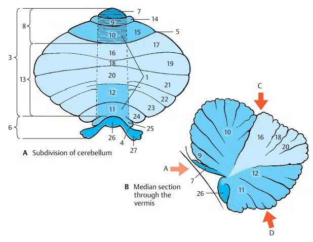
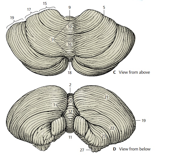
The cerebellum is the integrative organ for the coordination and fine-tuning of move-ment and for the regulation of muscle tone. Itdevelops from the alar plate of the brain stem and forms the roof of the fourth ven-tricle. The superior surface (C) is covered by the cerebrum. Embedded into theinferiorsurface (D) is the medulla oblongata (seep. 101, C). There is an unpaired central part, the vermis of the cerebellum (ACD1, B), and the twocerebellar hemispheres. This triparti-tion is only visible at the inferior surface, where the vermis forms the floor of a fossa, the vallecula of the cerebellum (D2). The sur-face of the cerebellum exhibits numerous narrow, almost parallel convolutions, the folia of the cerebellum.
Phylogenetic studies indicate that the cere-bellum consists of old portions (developed early in evolution, present in all vertebrates) and new portions (developed late, present only in mammals). Accordingly, the cerebel-lum is subdivided into two parts, the floc-culonodular lobe and the cerebellar body(A3).The two are separated by the posterolateralfissure (A4). The cerebellar body is furthersubdivided by the primary fossa (AC5) into anterior lobe andposterior lobe.
Flocculonodular Lobe (A6)
Together with the lingula (AB7), this is the oldest portion (archicerebellum). Function-ally, it is connected to the vestibular nuclei through its fiber tracts (vestibulocerebellum).
Anterior Lobe of the Cerebellar Body (A8)
This is a relatively old component; together with its central sections, which belong to the vermis (central lobule [A – C9], culmen [A – C10]) and other sections of the vermis (uvula [ABD11], pyramid [ABD12]), it forms the paleocerebellum. It receives the spinocerebellar tracts for proprioceptive sensibility from the muscles (spinocerebel-lum).
(A13)
This is the new portion (neocerebellum); its enormous enlargement in primates con-tributes significantly to the formation of the cerebellar hemispheres. It receives the large corticocerebellar tracts from the cerebral cortex via the pontine nuclei (pontocerebel-lum) and represents the apparatus for fine-tuning of voluntary movements.
Traditional Nomenclature
The individual sections of the cerebellum have traditional names unrelated to their development or function. According to this classification, most sections of the vermis are associated with a pair of hemispheric lobes: the central lobule (A – C9) with the wing of the central lobule (A14) on each side,the culmen (A – C10) with the quadrangularlobule (AC15), the declive (A–C16) with the simple lobule (AC17), the folium (A–C18)with thesuperior semilunar lobule (ACD19), the tuber (ABD20) with the inferior semi-lunar lobule (AD21) and part of the gracile lobule (AD22), thepyramid (ABD12) withpart of the gracile lobule and the biventrallobule (AD23), the uvula (AB11) with the tonsilla (A24) and the paraflocculus(A25),and the nodulus (AB26) with the flocculus (AD27). Only the lingula (AB7) is not as-sociated with any lateral lobe.
The pale red arrow A in diagram B refers to the direction of viewing the anterior surface of the cerebellum as illustrated on p. 155, A.
Related Topics