Chapter: Microbiology and Immunology: Bacteriology: Pneumococusm
Streptococcus pneumonia
Pneumococusm
Introduction
Pneumococcus was earlier classified as Diplococcus pneumoniae. The bacterium has now been reclassified asStreptococcuspneumoniae due to its genetic similarities to Streptococcus. S. pneumoniae, however, differs from streptococci in its mor-phology (by having a specific polysaccharide capsule), bile solubility, and optochin sensitivity.
Streptococcus pneumonia
A lot of advances have been made toward the better under-standing of the pathogenesis, antibiotic resistance, and use of vaccines in pneumococcal infections caused by S. pneumoniae.
Properties of the Bacteria
◗ Morphology
S. pneumoniae shows the following morphological features:
· They are Gram-positive cocci measuring 0.5–1.25 m in diameter. Older cells decolorize rapidly and might appear Gram negative.In clinical specimens, they appear typically “lanceolate shaped” with one end pointed and the other end round. They are arranged in pairs (diplococci) with the broad ends in apposition to each other. In cultures, they usu-ally appear more rounded and are arranged in short chains (Color Photo 18).
· They are capsulated. A polysaccharide capsule completely envelops each pair of cocci. The capsule is visualized by stain-ing it directly with specific stains or by Indian ink negative staining or by Quellung reaction (Color Photo 19).
· They are nonsporing and nonmotile.
◗ Culture
S. pneumoniae is an aerobe and a facultative anaerobe. It grows at an optimum temperature of 37°C (range 25–42°C) and pH 7.8 (range 6.5–8.3). The growth is enhanced by the presence of 5–10% CO2. Pneumococci are fastidious. They grow only on an enriched media, such as blood agar or chocolate agar (supple- mented with blood products), which will supply nutrients, pH buffers, etc.
Blood agar: On blood agar, morphology of the colonies var-ies depending on the nature of the strain (whether capsulatedor noncapsulated), the type of incubation (whether aerobic or anaerobic), and the time of incubation:
· Colonies on blood agar in anaerobic incubation show beta-hemolysis (greenish discoloration), but show alpha-hemolysis in aerobic incubation (Color Photo 20).
· Capsulated strains after overnight incubation produce round and mucoid colonies measuring 1–3 mm in diam-eter. Some strains, e.g., type 3 S. pneumoniae (most virulent), produce copious quantities of capsular material and hence produce large mucoid colonies. Noncapsulated strains produce small and flat colonies.
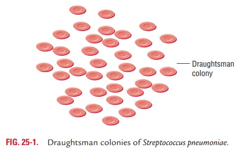
· On prolonged incubation, the colonies undergo autoly-sis and the centers become flattened or depressed (umbo-nation) and edges become raised, giving the colonies a typical draughtsman appearance (Fig. 25-1). The central flattening or depression is due to the production of intra-cellular enzymes, such as amidase, which lyses the bacteria. Bile salts, sodium lauryl sulfate, and other surface active agents enhance the process of autolysis of the bacterial colonies.

The capsules are present in strains isolated from clinical specimens but are lost on repeated cultivation, which is called smooth to rough variation.
Smooth to rough variation: The capsules are presentin strains isolated from clinical specimens but are lost on repeated cultivation. This is called smooth to rough varia-tion. Noncapsulated rough (R) strains are avirulent; these forms arise as spontaneous mutants and outgrow the parental smooth (S) strains in artificial culture. In tissues R forms are eliminated by phagocytosis. Transformation of a rough strain to a smooth one is also possible by treatment with smooth capsular substance containing DNA.
◗ Biochemical reactions
pneumoniae shows following reactions:
· S. pneumoniae ferments many sugars, producing acid onlybut no gas. Fermentation of sugars is carried out in Hiss’s serum water or in serum agar slopes.
· S. pneumoniae ferments inulin, and this is an important testto differentiate it from those streptococci that do not fer-ment inulin.
· S. pneumoniae produces an autolytic enzyme amidase, whichsolubilizes the peptidoglycan of the cell wall; hence in old cultures, typical draughtsman colonies are formed. This autolytic activity can be augmented by surface active agents, such as bile and bile salts.
· Bile solubility is a constant feature of pneumococci, and is positive in all the capsulated and some noncapsulated variants.
· Pneumococci are catalase and oxidase negative.
◗ Other properties
Susceptibility to physical and chemical agents: Pneumococciare delicate bacteria. They are killed by heating at 52°C for 15 minutes and by usual strengths of disinfectants. Pneumococcal colonies die on prolonged incubation.
Optochin sensitivity test:Pneumococciare sensitive to optochin (ethyl hydrocupreine)—a useful property to differentiate these from streptococci. Optochin sensitivity is usually performed by a paper disk containing 5 g of the compound; an inhibition zone of 14 mm or more indicates sensitivity to optochin. In recent times, occasional optochin-resistant pneumococci have also been documented. The target of optochin in pneumococci is an H ATPase, and resistance is thought to be due to a point mutation in one of subunit a or c of the H ATPase. This resistance to optochin is not related to the virulence of the bacteria.
Related Topics