Chapter: Essentials of Anatomy and Physiology: The Skeletal System
Skull - Skeleton
SKULL
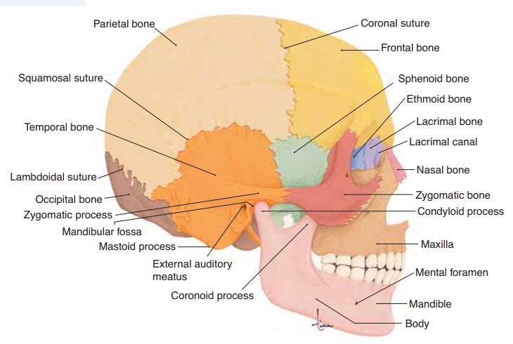
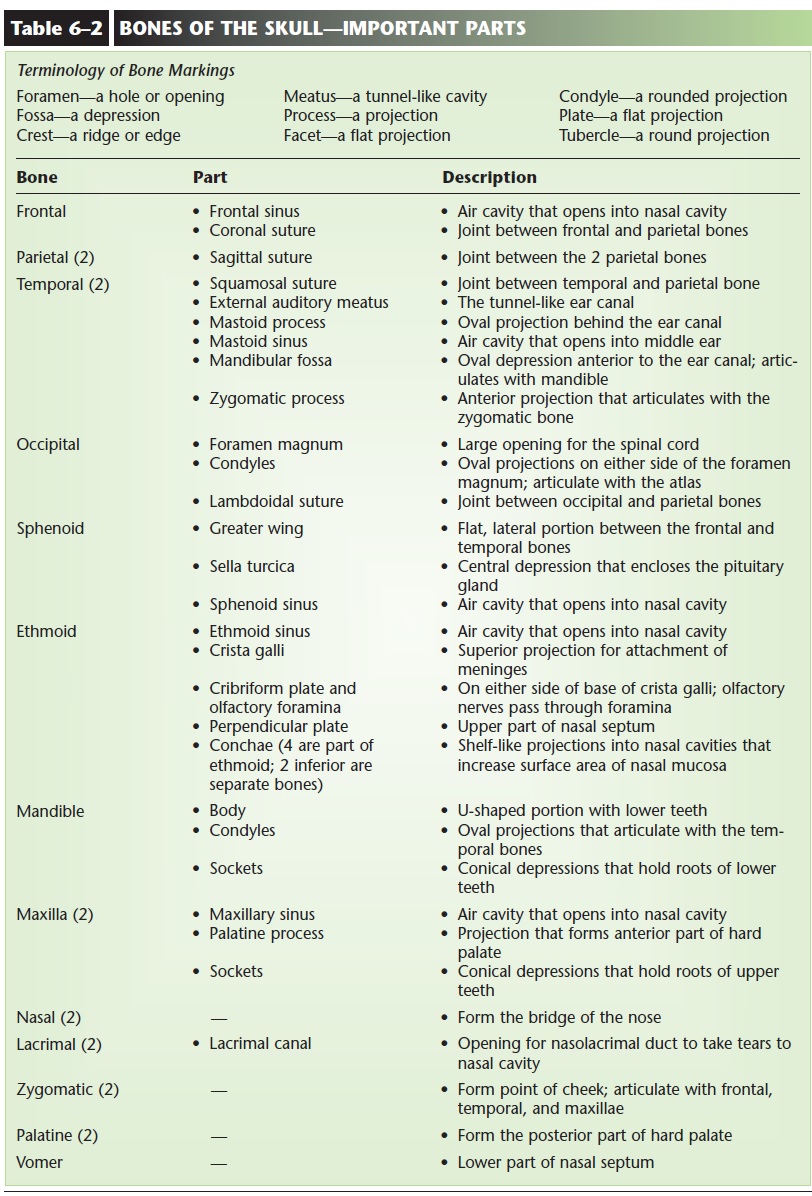
The skull consists of 8 cranial bones and 14 facial bones. Also in the head are three small bones in each middle ear cavity and the hyoid bone that supports the base of the tongue. The cranial bones form the brain-case (lined with the meninges) that encloses and pro-tects the brain, eyes, and ears. The names of some of these bones will be familiar to you; they are the same as the terminology used to describe areas of the head. These are the frontal bone, parietal bones (two), temporal bones (two), and occipital bone. The sphenoid bone and ethmoid bone are part of the floor of the braincase and the orbits (sockets) for the eyes. The frontal bone forms the forehead and the anterior part of the top of the skull. Parietal means “wall,” and the two large parietal bones form the posterior top and much of the side walls of the skull. Each temporal bone on the side of the skull contains an external auditory meatus (ear canal), a middle ear cav- ity, and an inner ear labyrinth. The occipital bone forms the lower, posterior part of the braincase. Its foramen magnum is a large opening for the spinal cord, and the two condyles (rounded projections) on either side articulate with the atlas, the first cervical vertebra. The sphenoid bone is said to be shaped like a bat, and the greater wing is visible on the side of the skull between the frontal and temporal bones. The body of the bat has a depression called the sella turcica, which encloses the pituitary gland. The ethmoid bone has a vertical projection called the crista galli (“rooster’s comb”) that anchors the cranial meninges. The rest of the ethmoid bone forms the roof and upper walls of the nasal cavities, and the upper part of the nasal septum.
All of the joints between cranial bones are immov-able joints called sutures. It may seem strange to refer to a joint without movement, but the term joint (or articulation) is used for any junction of two bones. In a suture, the serrated, or sawtooth, edges of adjacent bones fit into each other. These interlocking projections prevent sliding or shifting of the bones if the skull is subjected to a blow or pres-sure. In Fig. 6–5 you can see the coronal suture between the frontal and parietal bones, the squamosal suture between the parietal and temporal bones, and the lambdoidal suture between the occipital and pari-etal bones. Not visible is the sagittal suture, where the two parietal bones articulate along the midline of the top of the skull. All the bones of the skull, as well as the large sutures, are shown in Figs. 6–5 through 6–8. Their anatomically important parts are described in Table 6–2.
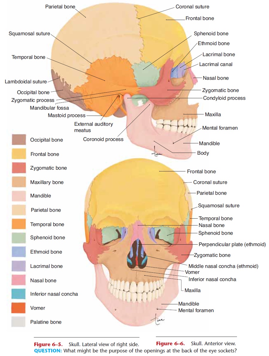
Figure 6–5. Skull. Lateral view of right side.
Figure 6–6. Skull. Anterior view.
QUESTION: What might be the purpose of the openings at the back of the eye sockets?
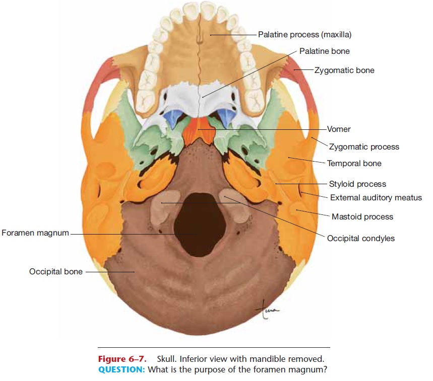
Figure 6–7. Skull. Inferior view with mandible removed. QUESTION: What is the purpose of the foramen magnum?
Of the 14 facial bones, only the mandible (lower jaw) is movable; it forms a condyloid joint with each temporal bone. The other joints between facial bones are all sutures. The maxillae are the two upper jaw bones, which also form the anterior portion of the hard palate (roof of the mouth). Sockets for the roots of the teeth are found in the maxillae and the mandible. The two nasal bones form the bridge of the nose where they articulate with the frontal bone (the rest of the nose is supported by cartilage). There is a lacrimal bone at the medial side of each orbit; the lacrimal canal contains the lacrimal sac, a passageway for tears. Each of the two zygomatic bones forms the point of a cheek, and articulates with the maxilla, frontal bone, and temporal bone. The two palatine bones are the posterior portion of the hard palate.
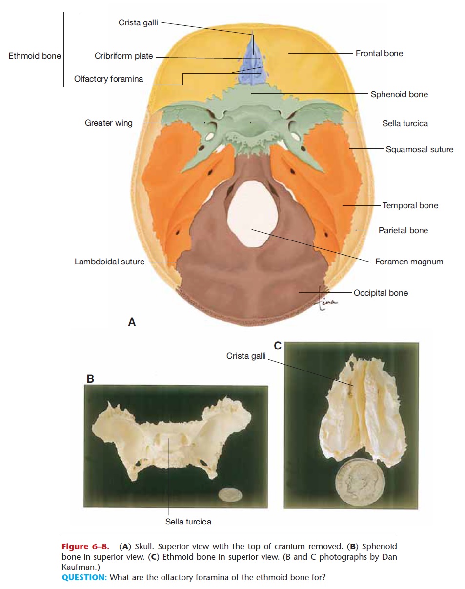
Figure 6–8. (A) Skull. Superior view with the top of cranium removed. (B) Sphenoid bone in superior view. (C) Ethmoid bone in superior view. (B and C photographs by Dan Kaufman.)
QUESTION: What are the olfactory foramina of the ethmoid bone for?
The plow-shaped vomer forms the lower part of the nasal septum; it articulates with the ethmoid bone. On either side of the vomer are the conchae, six scroll-like bones that curl downward from the sides of the nasal cavities; they help increase the surface area of the nasal mucosa. These facial bones are included in Table 6–2.
Paranasal sinuses are air cavities located in the maxillae and frontal, sphenoid, and ethmoid bones (Fig. 6–9). As the name paranasalsuggests, they open into the nasal cavities and are lined with ciliated epithelium continuous with the mucosa of the nasal cavities. We are aware of our sinuses only when they become “stuffed up,” which means that the mucus they produce cannot drain into the nasal cavities. This may happen during upper respiratory infections such as colds, or with allergies such as hay fever. These sinuses, however, do have functions: They make the skull lighter in weight, because air is lighter than bone, and they provide resonance for the voice, meaning more air to vibrate and thus deepen the pitch of the voice.
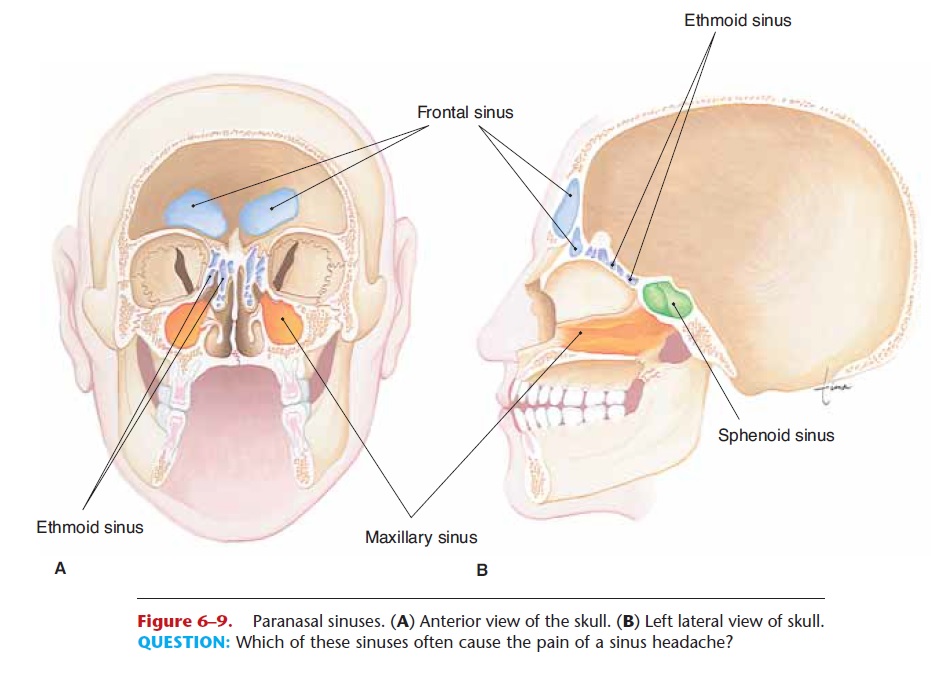
Figure 6–9. Paranasal sinuses. (A) Anterior view of the skull. (B) Left lateral view of skull. QUESTION: Which of these sinuses often cause the pain of a sinus headache?
The mastoid sinuses are air cavities in the mastoid process of each temporal bone; they open into the middle ear. Before the availability of antibiotics, mid-dle ear infections often caused mastoiditis, infection of these sinuses.
Within each middle ear cavity are three auditory bones: the malleus, incus, and stapes. As part of the hearing process, these bones transmit vibrations from the eardrum to the receptors in the inner ear (see Fig. 9–7).
Related Topics