Chapter: Nervous System and Sensory Organs : The Nervous System : An Overall View
Position of the Nervous System in the Body
Position of the Nervous System in the Body
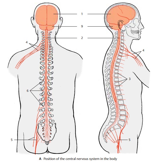
The central nervous system (CNS) is divided
into the brain, encephalon (A1), and the spinal cord (SC), medulla spinalis (A2). The brain in the cranial cavity is surrounded by a bony
capsule; the spinal cord in the vertebral canal is enclosed by the bony
vertebral column. Both are covered by meninges that enclose a cavity filled
with a fluid, the cerebrospinal fluid.
Thus, the CNS is protected from all sides by bony walls and the cushioning
effect of a fluid (fluid cush-ion).
The peripheral nervous system (PNS)
in-cludes the cranial nerves, which
emerge through holes (foramina) in
the base of the skull, and the spinal
nerves, which emerge through spaces between the vertebrae (in-tervertebral foramina) (A3). The peripheralnerves extend to
muscles and skin areas. They form nerve
plexuses before entering the limbs: the brachial plexus (A4) and
the lumbosacral plexus (A5) in which the fibers ofthe spinal
nerves intermingle; as a result, the nerves of the limbs contain portions of
different spinal nerves (see pp. 70 and 86). At the entry points of the
afferent nerve fibers lieganglia (A6); these are small oval bodies
containing sensory neurons.
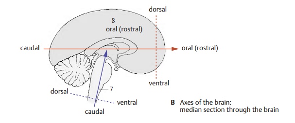
When
describing brain structures, terms like “top,” “bottom,” “front,” and “back”
are inaccurate, because we have to distinguish between different axes of the brain (B). Owing to the upright posture of humans, the neural tube is
bent; the axis of the spinal cord runs almost vertically, while the axis of the
forebrain (Forel’s axis, orange)
runs hori-zontally; the axis of the lower brain divi-sions (Meinert’s axis, violet) runs obliquely.
The positional terms relate to theses axes: the anterior end of the axis is
called oral or rostral (os, mouth; rostrum, beak), the pos-terior end is
called caudal (cauda, tail), the underside is called basal or ventral (venter,
abdomen), and the upper side is called dor-sal
(dorsum, back).
The
lower brain divisions, which merge
into the spinal cord, are collectively called the brain stem (light gray) (B7).
The anterior division is called the forebrain
(gray) (B8).
The
divisions of the brain stem, or encephalictrunk,
have a common structural plan (con-sisting of basal plate and alar plate,
like the spinal cord, see p. 13, C). Genuine peripheralnerves emerge from these divisions, as theydo from the
spinal cord. Like the spinal cord, they are supported by the chorda dorsalis during embryonic
development. All these features distinguish the brain stem from the forebrain.
The subdivision chosen here differs from the other classifications in which the
diencephalon is viewed as part of the brain stem.
The
forebrain, prosencephalon, consists
of two parts, the diencephalon and
the telen-cephalon or cerebrum. In the mature brain,the
telencephalon forms the two hemi-spheres (cerebral
hemispheres). The dien-cephalon lies between the two hemi-spheres.
A9
Cerebellum.
Related Topics