Chapter: Ophthalmology: Orbital Cavity
Orbital Cavity
Orbital Cavity
Basic Knowledge
Importance of the orbital cavity for the eye:
The orbital cavity is theprotec-tive bony socket for the globe together with the optic
nerve, ocular muscles,nerves, blood vessels, and lacrimal gland. These
structures are surrounded by orbital fatty tissue. The orbital cavity is shaped
like a funnel that opens anteri-orly
and inferiorly. The six ocular muscles originate at the apex of the funnel
around the optic nerve and insert into the globe. The globe moves within the
orbital cavity as in a joint socket.
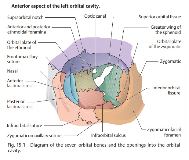
Bony socket:
This consists of seven bones (Fig. 15.1):
❖ Frontal.
❖ Ethmoid.
❖ Lacrimal.
❖ Sphenoid.
❖ Maxillary.
❖ Palatine.
❖ Zygomatic.

The bony rim of the orbital cavity forms a
strong ring. Its other bony surfaces include very thin plates of bone (see
adjacent structures).
Adjacent structures:
The close proximity of the orbital cavity to adjacentstructures
is clinically significant. The maxillary
sinus inferior to the orbital cavity is separated from it by a plate of
bone 0.5 mm thick. The ethmoidalair
cells located medial and posterior to the orbital cavity are separatedfrom
it by a plate of bone only 0.3 mm thick or
by periosteum alone. The fol-lowing other structures are also located immediately adjacent to the orbital
cavity.
❖ Sphenoidal sinus.
❖ Middle cranial fossa.
❖ Region of the optic chiasm.
❖ Pituitary gland.
❖ Cavernous sinus.
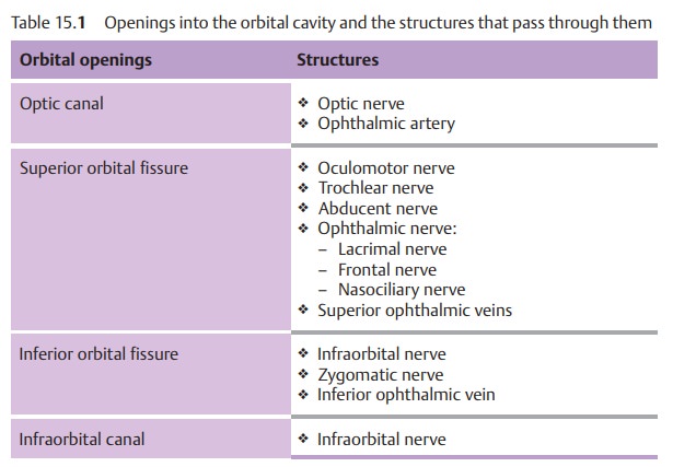
Superior adjacent structures include the anterior cranial fossa and the fron-tal sinus. Table 15.1 lists the various bony openings into the orbital cavity and the anatomic structures that pass through them.
Because of this anatomic sit-uation, the orbital cavity is frequently
affected by disorders of adjacent struc-tures. For example, inflammations of
the paranasal sinuses can result in orbi-tal cellulitis.
The walls of the orbital cavity are lined with
periosteum, which is also referred to as periorbita. Its anterior boundary is
formed by the orbital septa extending from the orbital rim to the superior and
inferior tarsal plates, the lateral and medial palpebral ligaments, and the
eyelids.
Arterial supply:
The orbital cavity is supplied by theophthalmic artery, abranch of the internal carotid artery. The
ophthalmic artery communicates with the angular artery, a branch of the
external carotid artery, via the supraorbital and supratrochlear arteries.
Stenosis of the internal carotid artery can result in reversed blood flow through the supraorbital and supratrochlear arteries. This can be dem-onstrated by Doppler ultrasound studies.
Venous drainage from the orbital cavity:
The orbital cavity drains throughthe inferior ophthalmic vein into the
pterygoid plexus, through the superiorophthalmic
vein into the cavernous sinus, and through the angular vein intothe facial veins.
Related Topics