Chapter: Medical Physiology: The Eye: III. Central Neurophysiology of Vision
Neuronal Patterns of Stimulation During Analysis of the Visual Image
Neuronal Patterns of Stimulation During Analysis of the Visual Image
Analysis of Contrasts in the Visual Image. If a person looksat a blank wall, only a few neurons in the primary visual cortex will be stimulated, regardless of whether the illumination of the wall is bright or weak. There-fore, the question must be asked, What does the primary visual cortex detect? To answer this, let us now place on the wall a large solid cross, as shown to the left in Figure 51–5. To the right is shown the spatial pattern of the most excited neurons in the visual cortex. Note that the areas of maximum excitationoccur along the sharp borders of the visual pattern.
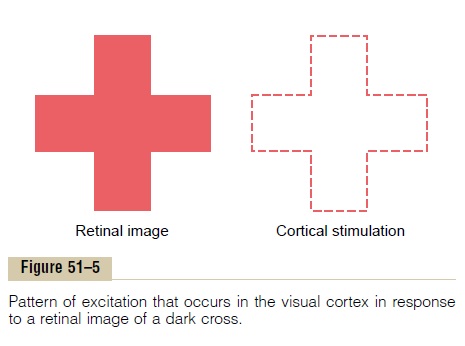
Thus, the visual signal in the primary visual cortex is concerned mainly with contrasts in the visual scene, rather than with noncontrasting areas. We saw that this is true of most of the retinal gan-glion cells as well, because equally stimulated adjacent retinal receptors mutually inhibit one another. But at any border in the visual scene where there is a change from dark to light or light to dark, mutual inhibition does not occur, and the intensity of stimulation of most neurons is proportional to the gradient of contrast— that is, the greater the sharpness of contrast and the greater the intensity difference between light and dark areas, the greater the degree of stimulation.
Visual Cortex Also Detects Orientation of Lines and Borders— “Simple” Cells
The visual cortex detects not only theexistence of lines and borders in the different areas of the retinal image but also the direction of orientation of each line or border—that is, whether it is vertical or horizontal or lies at some degree of inclination. This is believed to result from linear organizations of mutu-ally inhibiting cells that excite second-order neurons when inhibition occurs all along a line of cells where there is a contrast edge. Thus, for each such orienta-tion of a line, specific neuronal cells are stimulated. A line oriented in a different direction excites a different set of cells. These neuronal cells are called simple cells. They are found mainly in layer IV of the primary visual cortex.
Detection of Line Orientation When a Line Is Displaced Later-ally or Vertically in the Visual Field—“Complex” Cells. Asthe visual signal progresses farther away from layer IV, some neurons respond to lines that are oriented in the same direction but are not position-specific. That is, even if a line is displaced moderate distances laterally or vertically in the field, the same few neurons will still be stimulated if the line has the same direction. These cells are called complex cells.
Detection of Lines of Specific Lengths, Angles, or Other Shapes
Some neurons in the outer layers of the primary visual columns, as well as neurons in some secondary visual areas, are stimulated only by lines or borders of spe-cific lengths, by specific angulated shapes, or by images that have other characteristics. That is, these neurons detect still higher orders of information from the visual scene. Thus, as one goes farther into the analyt-ical pathway of the visual cortex, progressively more characteristics of each visual scene are deciphered.
Detection of Color
Color is detected in much the same way that lines are detected: by means of color contrast. For instance, a red area is often contrasted against a green area, a blue area against a red area, or a green area against a yellow area. All these colors can also be contrasted against a white area within the visual scene. In fact, this con-trasting against white is believed to be mainly respon-sible for the phenomenon called “color constancy”; that is, when the color of an illuminating light changes, the color of the “white” changes with the light, and appropriate computation in the brain allows red to be interpreted as red even though the illuminating light has changed the color entering the eyes.
The mechanism of color contrast analysis depends on the fact that contrasting colors, called “opponent colors,” excite specific neuronal cells. It is presumed that the initial details of color contrast are detected by simple cells, whereas more complex contrasts are detected by complex and hypercomplex cells.
Effect of Removing the Primary Visual Cortex
Removal of the primary visual cortex in the human being causes loss of conscious vision, that is, blindness.
However, psychological studies demonstrate that such “blind” people can still, at times, react subconsciously to changes in light intensity, to movement in the visual scene, or, rarely, even to some gross patterns of vision. These reactions include turning the eyes, turning the head, and avoidance. This vision is believed to be sub-served by neuronal pathways that pass from the optic tracts mainly into the superior colliculi and other por-tions of the older visual system.
Related Topics