Chapter: Human Nervous System and Sensory Organs : Autonomic Nervous System
Lower Thoracic and Abdominal Segments
Lower Thoracic and Abdominal Segments
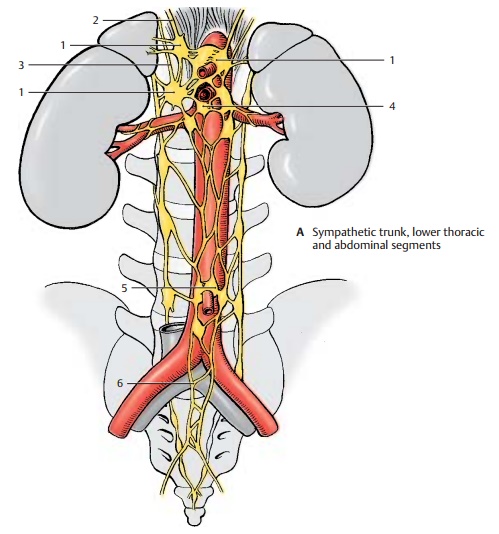
Branches originating from the thoracic and upper lumbar sympathetic trunk ganglia extend to the prevertebral ganglia of the abdominal aortic plexus. There are severalgroups of ganglia. At the exit of the celiac trunk lie the celiac ganglia (A1) where the greater splanchnic nerve (A2) (T5 – T9) andthe lesser splanchnic nerve (A3) (T9 – T11) terminate. Their postganglionic fibers ex-tend with the branches of the aorta to stom-ach, duodenum, liver, pancreas, spleen, and adrenal gland (gastric plexuses, hepatic plexus, splenic plexus, pancreatic plexus, suprarenal plexus). Preganglionic fibers run to the adrenal medulla .
The postganglionic fibers from the superiormesenteric ganglion (A4), together with thebranches of the celiac ganglion, supply the small intestine, the ascending colon, and the transverse colon. The fibers from the inferiormesenteric ganglion (A5) supply the de-scending colon, the sigmoid colon, and the rectum. The preganglionic fibers (lumbar splanchnic nerves) of both ganglia originate from levels T11 – L2. Some branches extend to the renal plexus, which also contains fibers from the celiac ganglia and the supe-rior hypogastric plexus.
Parasympathetic fibers also participate in the formation of the visceral plexuses. Stimulation of parasympathetic fibers in the digestive tract leads to increased peristalsis and secretion as well as relaxation of the sphincter muscles, while stimulation of sympathetic fibers causes reduced peristal-sic and secretion as well as contraction of the sphincter muscles.

The pelvic organs are supplied by the supe-rior hypogastric plexus (A6) and the inferior hypogastric plexus. Both plexuses receivepreganglionic sympathetic fibers from the lower thoracic and upper lumbar spinal cord, and parasympathetic fibers from the sacral spinal cord.
The urinary bladder is predominantly inner-vated by the parasympathetic fibers of the visceral plexus that supply the muscles for bladder contraction (detrusor muscle). The sympathetic fibers terminate at the smooth muscles of the orifice of the urethra and both ureteral orifices. Regulation of bladder tone and urination takes place via spinal re-flexes which, in turn, are controlled by the hypothalamus and by cortical areas.
The genitals are supplied by the prostaticplexus in the male and by the uterovaginal plexus in the female. Stimulation of para-sympathetic fibers dilates the vessels of the cavernous bodies and thus triggers erection in the male (nervi erigentes). Stimulation of sympathetic fibers leads to vasoconstriction and to ejaculation. The uterine muscles are innervated by sympathetic and parasympa-thetic fibers. Their functional significance is not clear because even a denervated uterus is fully functional during pregnancy and parturition.
Related Topics