Chapter: Basic Radiology : Plain Film of the Abdomen
Exercise: Increased Abdominal Density or Masses
EXERCISE 8-3.
INCREASED ABDOMINAL DENSITY OR MASSES
8-9. What is the most
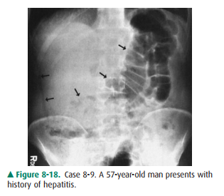
likely diagnosis of this soft-tissue mass (arrows) in Case 8-9 (Figure 8-18)?
A.
Ascites
B.
Cirrhosis
C.
Hepatomegaly
D.
Nephromegaly
8-10. What is the most
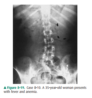
likely diagnosis of this soft-tissue mass (arrows) in Case 8-10 (Figure 8-19)?
A.
Adrenal carcinoma
B.
Gastric outlet obstruction
C.
Renal cell carcinoma
D.
Splenomegaly
8-11. What is the most
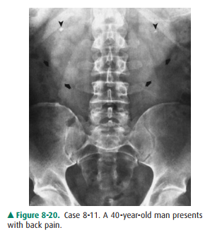
likely diagnosis of this soft-tissue mass (arrows) in Case 8-11 (Figure 8-20)?
A.
A pseudotumor sign of small bowel obstruction
B.
Gastric outlet obstruction
C.
Hepatomegaly
D.
Horseshoe kidney
8-12. What is the most
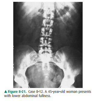
likely diagnosis in Case 8-12 (Figure8-21)?
A.
Ovarian cyst
B.
Pelvic abscess
C.
Pelvic hematoma
D.
Pelvic kidney
Radiologic Findings
8-9. In this case, the right side of
the abdomen shows increased density
and is relatively
free of gas. Displacement of the gas pattern in the
duodenum and jejunum to the left side is indicative of hepatomegaly (C is the
correct answer to Question 8-9). Hepatic metastases from lung cancer were later
confirmed.
8-10. In this case, a
soft-tissue mass in the left upper quad-rant displaces the gas in the splenic
flexure of the colon downward. Left adrenal or renal cell carcinoma rarely
presents as a large mass to the left of the mid-line. The most likely diagnosis
is splenomegaly (D is the correct answer to Question 8-10).
8-11. In this case, a
mass in the midabdomen delineates the lower poles of both kidneys, which are
fused at the midline, consistent with horseshoe kidney (D is the correct answer
to Question 8-11). Small renal cal-culi (arrowheads) are present bilaterally.
8-12. This case shows a
soft-tissue mass in the pelvis. In a middle-aged woman, an ovarian or uterine
mass would be the most likely considerations. Ultrasonog-raphy of the pelvis
showed a large, fluid-filled mass, confirmed surgically as an ovarian cyst (A
is the cor-rect answer to Question 8-12).
Discussion
Although abdominal plain
radiographs are useful in detecting hepatomegaly or splenomegaly, they are of
little use in diag-nosing hepatic disease, particularly if hepatomegaly is not
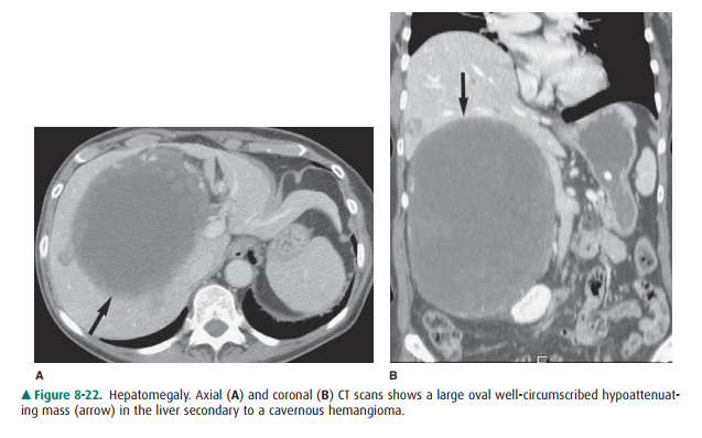
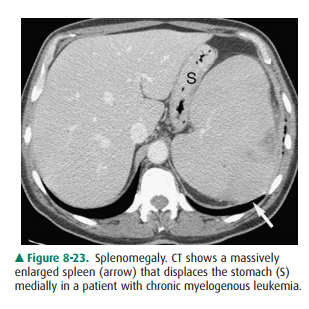
present. Other imaging modalities, such as ultrasonography, CT (Figures 8-22,
8-23), MR imaging, and radionuclide liver scans, are more sensitive and
accurate for evaluating hepatic primary diseases or metastases. In addition,
barium studies of the gastrointestinal tract and CT may be helpful in excluding
gastric outlet obstruction, carcinoma, or renal cell carcinoma.
Fusion of the kidneys may occur
in the embryologic stage during the second month of gestation. Most (95%) of
these fusions occur at the lower poles of the kidneys. CT shows the kidney to
be vertical or even in the reverse oblique directionand its position to be
lower than normal. Horseshoe kidney may be associated with other congenital
anomalies, as well as a high incidence of urinary tract obstruction, infection,
or stone formation. A horseshoe kidney may also deviate the upper ureters
laterally.
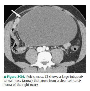
When the plain film suggests the
presence of a pelvic mass, a specific diagnosis is often not possible. Pelvic
ultra-sonography, CT (Figure 8-24), or MR imaging better demon-strates the
pelvic organs and their interrelationship and will differentiate between solid
and fluid content in the mass.
Related Topics