Chapter: Essentials of Anatomy and Physiology: Tissues and Membranes
Epithelial Tissue

EPITHELIAL TISSUE
Epithelial tissues are found on surfaces as either cov-erings (outer surfaces) or linings (inner surfaces). Because they have no capillaries of their own, epithe-lial tissues receive oxygen and nutrients from the blood supply of the connective tissue beneath them. Many epithelial tissues are capable of secretion and may be called glandular epithelium, or more simply, glands.
Classification of the epithelial tissues is based on the type of cell of which the tissue is made, its charac-teristic shape, and the number of layers of cells. There are three distinctive shapes: squamous cells are flat, cuboidal cells are cube shaped, and columnar cells are tall and narrow. “Simple” is the term for a single layer of cells, and “stratified” means that many layers of cells are present (Fig. 4–1).
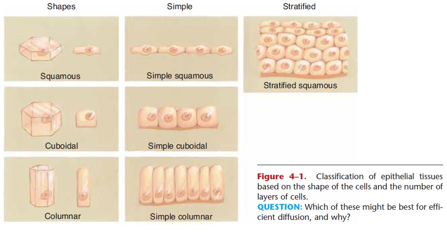
Figure 4–1. Classification of epithelial tissues based on the shape of the cells and the number of layers of cells.
QUESTION: Which of these might be best for effi-cient diffusion, and why?
SIMPLE SQUAMOUS EPITHELIUM
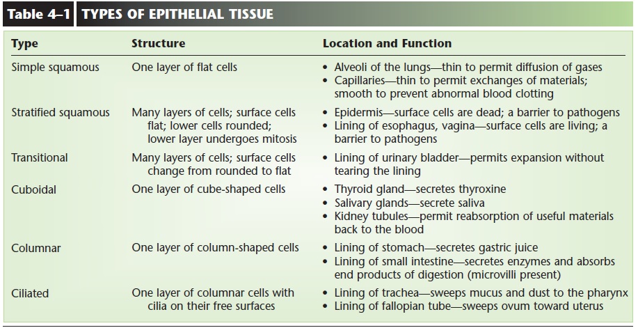
Simple squamous epithelium is a single layer of flat cells (Fig. 4–2). These cells are very thin and very smooth—these are important physical characteristics. The alveoli (air sacs) of the lungs are simple squamous epithelium. The thinness of the cells permits the dif-fusion of gases between the air and blood.
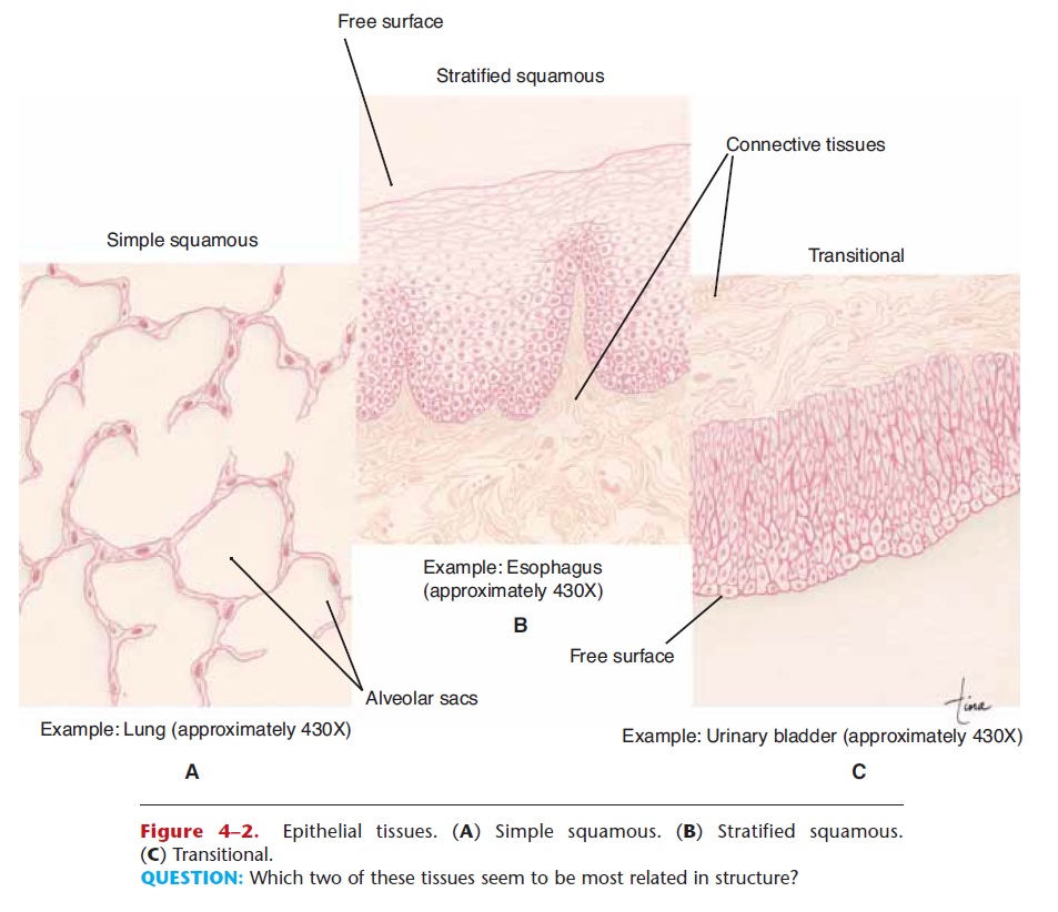
Figure 4–2. Epithelial tissues. (A) Simple squamous. (B) Stratified squamous. (C) Transitional.
QUESTION: Which two of these tissues seem to be most related in structure?
Another location of this tissue is capillaries, the smallest blood vessels. Capillary walls are only one cell thick, which permits the exchange of gases, nutrients, and waste products between the blood and tissue fluid. The interior surface of capillaries is also very smooth (and these cells continue as the lining of the arteries, veins, and heart); this is important because it prevents abnormal blood clotting within blood vessels.
STRATIFIED SQUAMOUS EPITHELIUM
Stratified squamous epithelium consists of many layers of mostly flat cells, although lower cells are rounded. Mitosis takes place in the lowest layer to continually produce new cells to replace those worn off the surface (see Fig. 4–2). This type of epithelium makes up the epidermis of the skin, where it is called “keratinizing” because the protein keratin is produced, and the surface cells are dead. Stratified squamous epithelium of the non-keratinizing type lines the oral cavity, the esophagus, and, in women, the vagina. In these locations the surface cells are living and make up the mucous membranes of these organs. In all of its body locations, this tissue is a barrier to microorgan-isms because the cells of which it is made are very close together.
TRANSITIONAL EPITHELIUM
Transitional epithelium is a type of stratified epithe-lium in which the surface cells change shape from round to squamous. The urinary bladder is lined with transitional epithelium. When the bladder is empty, the surface cells are rounded (see Fig. 4–2). As the bladder fills, these cells become flattened. Transitional epithelium enables the bladder to fill and stretch with-out tearing the lining.
SIMPLE CUBOIDAL EPITHELIUM
Simple cuboidal epithelium is a single layer of cube-shaped cells (Fig. 4–3). This type of tissue makes up the functional units of the thyroid gland and salivary glands. These are examples of glandular epithelium; their function is secretion. In these glands the cuboidal cells are arranged in small spheres and secrete into the cavity formed by the sphere. In the thyroid gland, the cuboidal epithelium secretes the thyroid hormones; thyroxine is an example. In the salivary glands the cuboidal cells secrete saliva. Cuboidal epithelium also makes up portions of the kidney tubules. Here the cells have microvilli (see Fig. 1–1), and their function is the reabsorption of useful materi-als back to the blood.
Figure 4–3. Epithelial tissues. (A) Simple cuboidal. (B) Simple columnar. (C) Ciliated. QUESTION: What is the function of the cilia that line the tracheaepithelium also makes up portions of the kidney tubules. Here the cells have microvilli (see Fig. 1–1), and their function is the reabsorption of useful materi-als back to the blood.
GLANDS
Glands are cells or organs that secrete something; that is, they produce a substance that has a function either at that site or at a more distant site.
Unicellular Glands
Unicellular means “one cell.” Goblet cells are an example of unicellular glands. As mentioned earlier, goblet cells are found in the lining of the respiratory and digestive tracts. Their secretion is mucus.
Multicellular Glands
Most glands are made of many similar cells, or of avariety of cells with their secretions mingled into acollective secretion. Multicellular glands may be divided into two major groups: exocrine glands and endocrine glands.
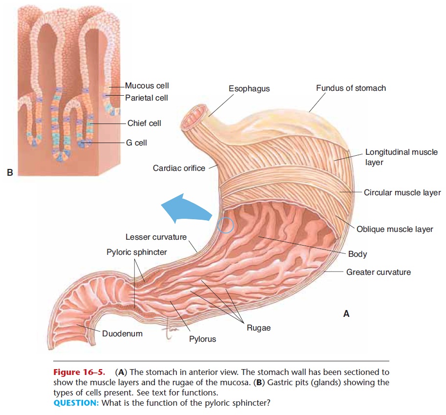
Exocrine glands have ducts (tubes) to take the secretion away from the gland to the site of its func-tion. Salivary glands, for example, secrete saliva that is carried by ducts to the oral cavity. Sweat glands secrete sweat that is transported by ducts to the skin surface, where it can be evaporated by excess body heat. Thegastric glands of the stomach lining contain different kinds of cells (see Fig. 16–5), which produce hydrochloric acid and the enzyme pepsin. Both of these secretions are part of gastric juice.
Endocrine glands are ductless glands. The secre-tions of endocrine glands are a group of chemicals called hormones, which enter capillaries and are cir-culated throughout the body. Hormones then bring about specific effects in their target organs. These effects include aspects of growth, use of minerals and other nutrients, and regulation of blood pressure, and will be covered in more detail. Examples of endocrine glands are the thyroid gland, adrenal glands, and pituitary gland.
The pancreas is an organ that is both an exocrine and an endocrine gland. The exocrine portions secrete digestive enzymes that are carried by ducts to the duodenum of the small intestine, their site of action. The endocrine portions of the pancreas, called pan-creatic islets or islets of Langerhans, secrete the hor-mones insulin and glucagon directly into the blood.
Related Topics