Chapter: 12th Zoology : Chapter 7 : Human Health and Diseases
Common diseases in human beings
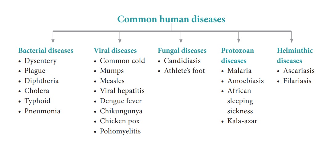
Common
diseases in human beings
Disease can be defined
as a disorder or malfunction of the mind or body. It involves morphological,
physiological and psychological disturbances which may be due to environmental
factors or pathogens or genetic anomalies or life style changes. Diseases can
be broadly grouped into infectious and non infectious types.
Diseases which are transmitted from one person to another are called infectious diseases or communicable diseases. Such disease causing organisms are called pathogens and are transmitted through air, water, food, physical contact and vectors. The disease causing pathogen may be virus, bacteria, fungi, protozoan parasites, helminthic parasites, etc., Infectious diseases are common and everyone suffers from such diseases at some time or the other. Most of the bacterial diseases are curable but all viral diseases are not. Some infectious disease like AIDS may be fatal.
Non-infectious diseases are not transmitted
from an infected person to a healthy person. In origin they may be genetic
(cystic fibrosis), nutritional (vitamin deficiency diseases) and degenerative
(arthritis, heart attack, stroke). Among non - infectious diseases, cancer is
one of the major causes of death.
1. Bacterial and viral diseases
Bacterial diseases
Though the number of
bacterial species is very high, only a few bacteria are associated with human
diseases and are called pathogenic bacteria. Such pathogens may emit
toxins and affected the body. Common pathogenic bacteria and the bacterial
diseases are given in table 7.1.
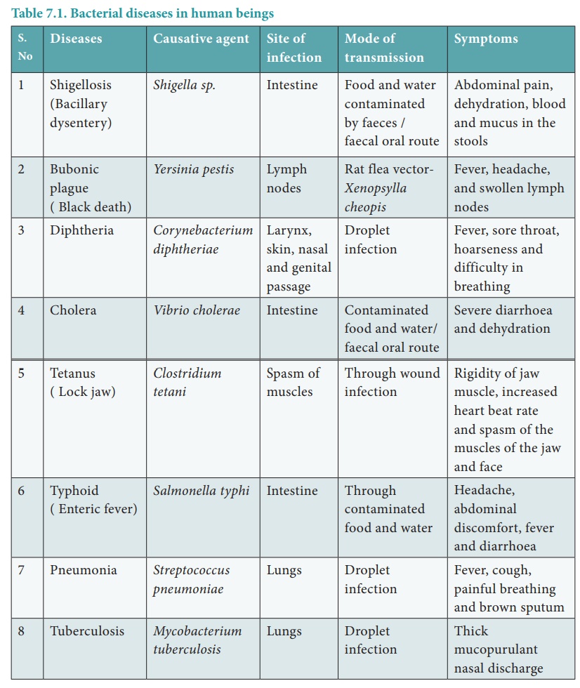
Bacteria spread through
air, water or by inhaling the droplets/aerosols or even by sharing utensils,
dresses with an infected person. Typhoid fever can be confirmed by Widal
test.
Viral
diseases
Viruses are the smallest intracellular obligate parasites, which multiply within living cells. Outside the living cells they cannot carry out the characteristics of a living organism. Viruses invade living cells, forcing the cells to create new viruses. The new viruses break out of the cell, killing it and invade other cells in the body, causing diseases in human beings. Rhino viruses cause one of the most infectious human ailment called the “Common cold”.
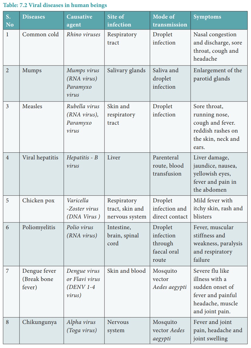
Viral diseases are
generally grouped into four types on the basis of the symptoms produced in the
body organs.
i.
Pneumotropic diseases (respiratory tract infected by influenza)
ii. Dermotropic diseases (skin and subcutaneous tissues affected by chicken pox and measles)
iii.
Viscerotropic diseases (blood and visceral organs affected by
yellow fever and dengue fever)
iv.
Neurotropic diseases (central nervous system affected by rabies
and polio). Some common viral diseases of human beings are given in table
7.2.
2. Protozoan diseases
About 15 genera of
protozoans live as parasites within the human body and cause diseases.
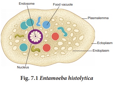
Amoebiasis also called amoebic
dysentery or amoebic colitis is caused by Entamoeba histolytica,
which lives in the human large intestine and feeds on food particles
and bacteria (Fig. 7.1). Infective stage of this parasite is the trophozoite,
which penetrates the walls of the host intestine (colon) and secretes
histolytic enzymes causing ulceration, bleeding, abdominal pain and stools with
excess mucus. Symptoms of amoebiasis can range from diarrhoea to dysentery with
blood and mucus in the stool. House flies (Musca domestica)
acts as a carrier for transmitting the parasite from contaminated
faeces and water.
African sleeping
sickness is
caused by Trypanosoma species. Trypanosoma is
generally transmitted by the blood sucking Tsetse flies. Three species
of Trypanosoma cause sleeping sickness in man.
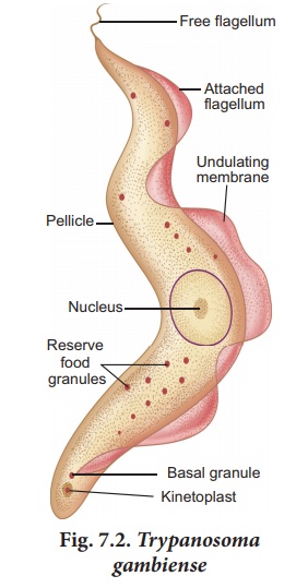
1.
T. gambiense is transmitted by Glossina palpalis (Tsetse fly) and
causes Gambian or Central African sleeping sickness (Fig. 7.2).
2.
T. rhodesiense is transmitted by Glossina morsitans causing
Rhodesian or East African sleeping sickness.
3.
T. cruzi is transmitted by a bug called Triatoma megista and causes
Chagas disease or American trypanosomiasis.
Kala – azar or visceral
leishmaniasis is caused by Leishmania donovani, which is
transmitted by the vector Phlebotomus (sand fly).
Infection may occur in the endothelial cells, bone marrow, liver, lymph
glands and blood vessels of the spleen. Symptoms of Kala azar are weight loss,
anaemia, fever, enlargement of spleen and liver.
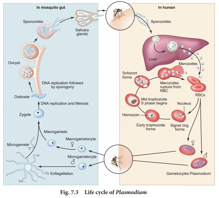
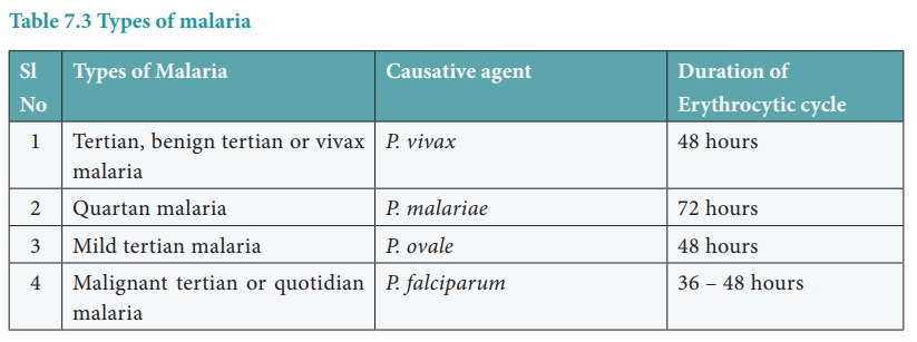
Malaria is caused by different
types of Plasmodium species such as P. vivax, P. ovale, P.
malariae and P. falciparum (Table 7.3). Plasmodium lives in
the RBC of human in its mature condition it is called as trophozoite.
It is transmited from one person to another by the bite of the infected female Anopheles
mosquito.
Life
cycle of Plasmodium
Plasmodium vivax is a digenic
parasite, involving two hosts, man as the secondary host and female Anopheles
mosquito as the primary host. The life cycle of Plasmodium
involves three phases namely schizogony, gamogony and sporogony (Fig.
7.3).
The parasite first enters
the human blood stream through the bite of an infected female Anopheles
mosquito. As it feeds, the mosquito injects the saliva
containing the sporozoites. The sporozoite within the blood stream
immediately enters the hepatic cells of the liver. Further in the liver they
undergo multiple asexual fission (schizogony) and produce merozoites.
After being released from liver cells, the merozoites penetrate the RBC’s.
Inside the RBC, the
merozoite begins to develop as unicellular trophozoites. The trophozoite grows
in size and a central vacuole develops pushing them to one side of cytoplasm
and becomes the signet ring stage. The trophozoite nucleus then divides
asexually to produce the schizont. The large schizont shows yellowish -
brown pigmented granules called Schuffners granules. The schizont
divides and produces mononucleated merozoites. Eventually the
erythrocyte lyses, releasing the merozoites and haemozoin toxin into the blood
stream to infect other erythrocytes. Lysis of red blood cells results in cycles
of fever and other symptoms. This erythrocytic stage is cyclic and repeats
itself approximately every 48 to 72 hours or longer depending on
the species of Plasmodium involved. The sudden release of
merozoites triggers an attack on the RBCs. Occasionally, merozoites
differentiate into macrogametocytes and microgametocytes. When
these are ingested by a mosquito, they develop into male and female gametes
respectively.
In the mosquito's gut,
the infected erythrocytes lyse and male and female gametes fertilize to form a
diploid zygote called ookinete. The ookinete migrates to the mosquito's
gut wall and develop into an oocyte. The oocyte undergoes meiosis by a
process called sporogony to form sporozoites. These sporozoites
migrate to the salivary glands of the mosquito. The cycle is now completed and
when the mosquito bites another human host, the sporozoites are injected and
the cycle begins a new.
The pathological changes caused by malaria, affects not only the erythrocytes but also the spleen and other visceral organs. Incubation period of malaria is about 12 days. The early symptoms of malaria are headache, nausea and muscular pain.
The classic symptoms first develop with the synchronized release of merozoites, haemozoin toxin and erythrocyte debris into the blood stream resulting in malarial paroxysms – shivering chills, high fever followed by sweating. Fever and chills are caused partly by malarial toxins that induce macrophages to release tumour necrosis factor (TNF-α) and interleukin.
Prevention
It is possible to break the transmission cycle by killing the insect vector. Mosquito's lay their eggs in water. Larvae hatch and develop in water but breathe air by moving to the surface. Oil can be sprayed over the water surface, to make it impossible for mosquito larvae and pupae to breathe.
Ponds, drainage ditches
and other permanent bodies of water can be stocked with fishes such as Gambusia
which feed on mosquito larvae. Preparations containing Bacillus
thuringiensis can be sprayed to kill the mosquito larvae since it is
not toxic to other forms of life. The best protection against malaria is to
avoid being bitten by mosquito. People are advised to use mosquito nets, wire
gauging of windows and doors to prevent mosquito bites.
In the 1950’s the World
Health Organisation (WHO) introduced the Malaria eradication programme. This
programme was not successful due to the resistance of Plasmodium to the
drugs used to treat it and resistance of mosquito's to DDT and other
insecticides.
3. Fungal diseases
Fungi was recognized as
a causative agent of human diseases much earlier than bacteria. Dermatomycosis
is a cutaneous infection caused by fungi belonging to the genera Trichophyton,
Microsporum and Epidermophyton.
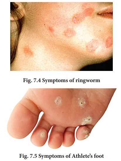
Ringworm is one of the most common fungal disease in humans (Fig. 7.4). Appearance of dry, scaly lesions on the skin, nails and scalp are the main symptoms of the disease. Heat and moisture help these fungi to grow and makes them to thrive in skin folds such as those in the groin or between the toes. Ringworms of the feet is known as Athlete’s foot caused by Tinea pedis (Fig. 7.5) . Ringworms are generally acquired from soil or by using clothes, towels and comb used by infected persons.
4. Helminthic diseases
Helminthes are mostly
endoparasitic in the gut and blood of human beings and cause diseases called helminthiasis.
The two most prevalent helminthic diseases are Ascariasis and Filariasis.
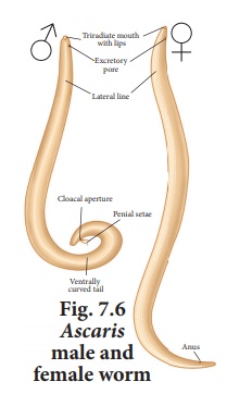
Ascaris is a monogenic parasite and exhibits sexual dimorphism. Ascariasis is a disease caused by the intestinal endoparasite Ascaris lumbricoides commonly called the round worms (Fig. 7.6). It is transmitted through ingestion of embryonated eggs through contaminated food and water. Children playing in contaminated soils are also prone to have a chance of transfer of eggs from hand to mouth.
The symptoms of the disease are abdominal pain,
vomiting, headache, anaemia, irritability and diarrhoea. A heavy infection can
cause nutritional deficiency and severe abdominal pain and causes stunted
growth in children. It may also cause enteritis, hepatitis and bronchitis.
Filariasis is caused by Wuchereria
bancrofti , commonly called filarial worm. It
is found in the lymph vessels and lymph nodes of man
(Fig. 7.7). Wuchereria bancrofti is sexually
dimorphic, viviparous and digenic. The life cycle is completed in two
hosts, man and the female Culex mosquito The female filarial worm gives
rise to juveniles called microfilariae larvae. In the lymph
glands, the juveniles develop into adults. The accumulation of the worms
block the lymphatic system resulting in inflammation of the lymph nodes.
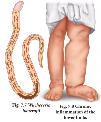
In some cases, the
obstruction of lymph vessels causes elephantiasis or filariasis of the limbs, scrotum and mammary glands (Fig. 7.8).
Related Topics