Chapter: 11th Zoology : Chapter 12 : Basic Medical Instruments and Techniques
Basic Biomedical Techniques
Biomedical Techniques
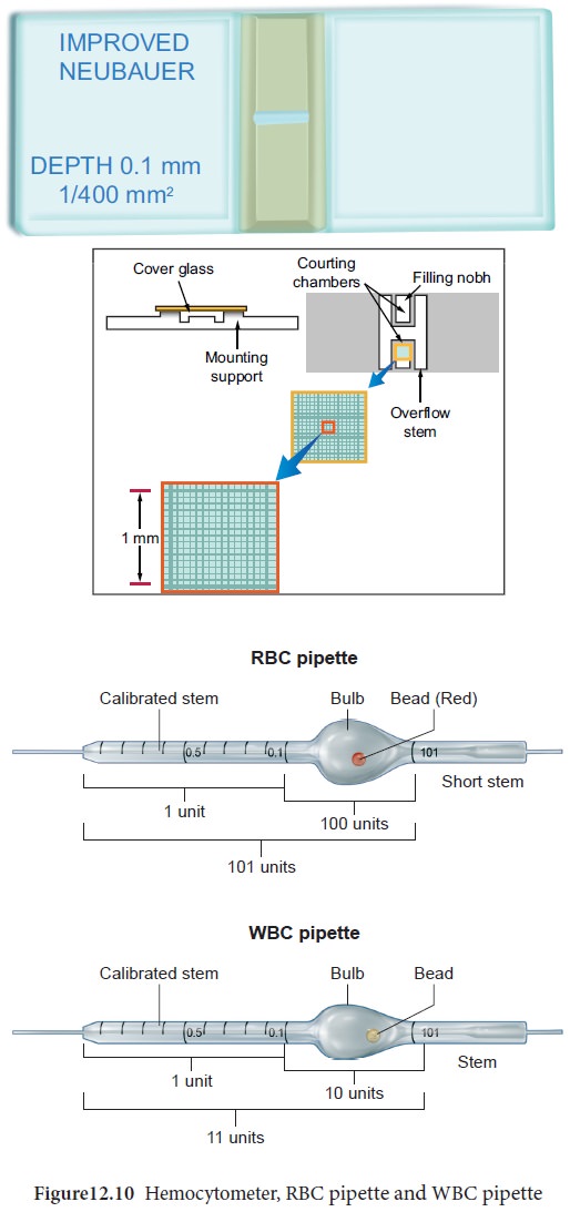
Blood Cell Counting using Haemocytometer
The haemocytometer is a thick glass slide with a
counting chamber in the middle. The counting chamber contains two grids with
improved Neubaur rulings of 3 by 3 mm primary square. The primary square is
further subdivide into 9 secondary squares, each 1 by 1 mm. The four corner
squares are used for the white blood cell count which are further subdivided
into 16 tertiary squares. The central secondary square is divided into
25 tertiary squares, each of which measure 0.2 by 0.2 mm, each single tertiary
square is further divided into 16 smaller squares. The five black squares along
with the shaded squares in the centre are used for platelet count, while the
five black squares alone are used for red blood cells counting (Figure12.10).
Diluting fluid
The blood cells are diluted in specific diluting fluid
to keep the cells intact. RBC diluting fluid (Hayem’s) is isotonic with blood,
hence haemolysis does not occur. The blood is diluted 1:200 times with RBC
diluting fluid and the cells are counted under 45X objective of the microscope.
The diluting fluid used for WBC count is Turk’s
solution which contains glacial acetic acid and Gentian violet. The glacial
acetic acid lyses the red blood cells and the Gentian violet stains the nuclei
of the leucocytes. The blood is diluted 1:20 times and the cell are counted
under 10X objective of the microscope. The total number of cells counted is
expressed in mm3.
Blood cell counting using hemocytometer
1. The blood
is collected till the 0.5 graduation in the pipette.
2. The
diluting fluid is taken till the graduations 11 and 101 of the WBC and RBC
pipette respectively.
3. The blood
is diluted and mixed well with the respective diluting fluid by rotating the
pipette horizontally several times.
4. The cover
slip is placed on top of the counting chamber.
5. The tip
of the pipette is placed on the counting chamber and fluid discharged till it
fills the chambers.
6. The cells
are allowed to settle for several minutes and the ruled area is viewed under
the microscope.
Preparation of Blood Smear
The examination of peripheral dry blood smear is a
very important laboratory test as it is possible to
• Estimate approximately the number of cellular components
• Study the morphology of these components
• Observe the presence of blood parasites
• Study the response of the body to various
diseases
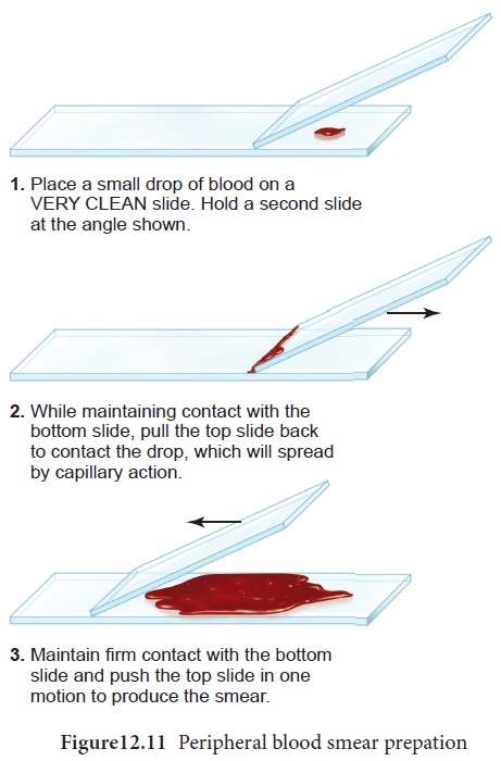
The methodology for the preparation of blood smear
is as follows (Figure12.11)
1. Place a
drop of blood on a clean glass slide about 1cm from one end
2. Using
another glass slide placed at an angle of about 45o to the previous slide.
3. Spread
the drop of blood quickly in one stroke as a thin film
4. Stain the
film using Leishman’s stain
5. Allow the
slide to dry and wash the excess stain
6. Observe
the slide under a light microscope (Figure12.12).
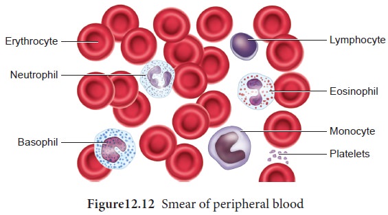
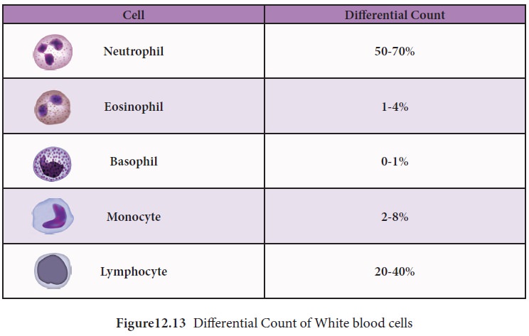
Differential Count
The differential WBC count is the method in which
the numbers of different types of white blood cells present in the blood are
counted by examining a well stained peripheral blood smear (prepared by the
above method). The number of each type of white blood cell is then expressed as
a percentage of the total number of cells counted (Figure12.13).
Related Topics