Chapter: Pharmaceutical Drug Analysis: Ultraviolet and Absorption Methods
Ultraviolet and Absorption Methods: Instrumentation
INSTRUMENTATION
A spectrophotometer is an instrument which is capable of
isolating ‘monochromatic’ radiation
; or that which specifically contains a dispersing element : a prism or a
grating.
It is pertinent to mention here that there are a plethora
of commercially available spectrophotometers of varying design i.e., single-beam (simple), double-beam
(more precise and accurate) and microcomputer controlled built-in-recorder with
separate printer ; and obviously having a wide-price-range from Rs 3.0 Lacs to
Rs 17.5 Lacs. Evidently, it is practically impossible to describe either all or
even a major fraction of, the various spectrophotometers available.
Therefore, in this particular section the following two types of spectrophotometers shall be
discussed briefly :
(a) Single-beam
Spectrophotometer, and
(b) Double-beam
Spectrophotometer.
1. SINGLE BEAM SPECTROPHOTOMETER
The desired wavelength is isolated by using a prism or
grating and auxiliary mirrors and slits that collectively from a microchromator
of the instrument. The wavelength dial on a spectrophotometer is adjusted to a
specific value, but the radiation leaving the exit-slit is found to be rarely
monochromatic. The schematic diagram of the Beckman Model DU-Spectrophotometer
is illustrated in Figure 21.5.
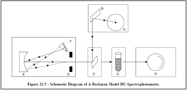
The various components of Figure 21.5 are given below :
A = Source of light ;
B = Condensing mirror ;
C = Slit-entrance mirror
;
D = Adjustable slit ;
E = Collimator mirror ;
F = Prism (Reflecting) ;
G = Cuvette containing
sample ;
H = Phototube ;
Light from the source (A) is focussed on the condensing
mirror (B) and directed in a beam to the 45° slit-entrance mirror (C). The
slit-entrance mirror subsequently deflects the beam through the adjustable slit
and into the monochromator to the collimator mirror (E). As a result the light
falling on the collimator mirror is rendered parallel and reflected to the
prism (F), where it undergoes refraction. The back surface of the prism is
aluminized, so that the light refracted at the first surface is reflected back
through the prism, undergoing further refraction as it emerges. The desired
wavelength of light is selected by rotating the wave-length selector fixed on
top of the monochromator case. This control, in fact, adjusts the position of
the prism. The spectrum from the prism is directed back to the collimating
mirror which centres the chosen wavelength of light on the slit and the sample
(G). Light passing through the sample strikes the phototube (H), causing a
voltage to appear across a load-resistor. The voltage is duly amplified and
registered on either the strip-chart recorder or the null-meter.
The Milton Roy
Spectronic(R)-20 definitely provides a low-cost and easy to
operate instrument, that is still capable of achieving absorbance readings
accurate to ± 1 or 2%.
Beckman Instruments, one of the pioneers in Analytical
Instruments and dominating this field since 50 years, has come up with their
latest Beckman DU Series 60
Spectrophotometer, which essentially makes use of two different sources of light, namely :
(a) H2
or D2 Lamp-for measurement in UV-region, and
(b) Tungsten
Lamp-for measurement in visible region,
thereby permitting measurements from 190-1000 nm. A
computer system has also been provided to enable automatic spectrochemical
measurements and perform calculations simultaneously.
2. DOUBLE BEAM SPECTROPHOTOMETER
The quantum leap amalgamated with qualified success in
the advancement of Analytical Instruments necessitated for more rapid and
precise and accurate measurements in UV and visible spectroscopy. It could be
accomplished by the help of the following two
cardinal modifications, namely :
(a) Need for a
continuous change in wavelength so that light through the blank and through the
sample may be monitored continuously, and
(b)
Measurements done with a recording spectrophotometer.
The above two modifications have been duly incorporated
in a double-beam spectrophotometer. Fig-ure 21.6, depicts the schematic diagram
of the optical system involved in a Lambda-2 microcomputer-con-trolled UV-VIS
Spectrophotometer (Perkin-Elmer).
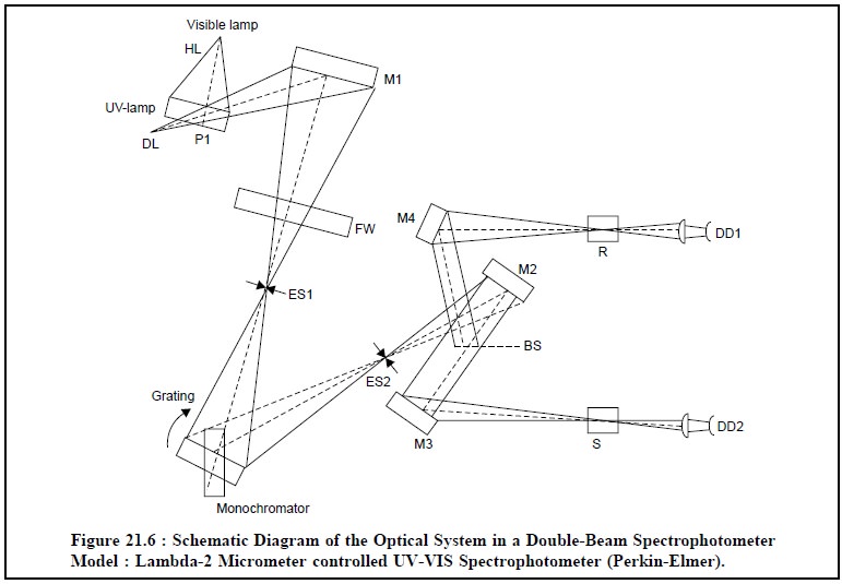
The various components of Figure 21.6 are stated below :
VIS-LAMP = Tungstem Lamp.
UV-LAMP = Hydrogen Lamp (HL),
= Deuterium Lamp (DL),
P1 = Movable source-selection mirror,
M1, M2, M3, M4
= Mirrors,
FW = Filter wheel,
ES 1 = Entrance slit,
ES 2 = Exit slit,
BS = Beam Splitter,
R = Reference Sample Holder,
S = Sample holder (Test), and
DD 1, DD 2 = Diode detectors.
In fact, the source beam is usually split in two
different manners, namely :
(a) Separated in Space : In this instance,
the source beam is split between the sample cell-path and the reference
cell-path, and finally detected by two diode detectors. Here, the two detectors
should be adequately matched so that no changes occur relative to each other
during the measurements,
(b) Separated in Time : In this case, the
source beam is split with the help of an optical chopper which permits the
source beam to alternate between the sample cell-path and the reference
cell-path. Here, the source should be stable enough so that no changes take
place in the radiant energy during the chopping time.
Keeping in view, this specific, rigid and stringent
requirement, the separation-in-space method is found to be normally of lower
precision and accuracy than the separation-in time-method.
Evidently, the optical choppers are quite expensive, and
therefore, the instrument manufacturers very often utilize the
separation-in-space method for the routine measurement spectrophotometers.
However, the most sophisticated double-beam
spectrophotometer is usually pretty expensive by vir-tue of the following
facts, namely :
(i) Greater
operating stability,
(ii) Rapid
speed compared to single-beam instruments,
(iii)
Complicated optical system involved, and
(iv) Recording
device for recording absorbance Vs wavelength.
The source beam after passing through the movable source
selection mirror (M1), gets reflected and subsequently makes an entry through
the filter wheel (FW) and the entrance-slit (ES 1) to the monochromator. The
grating is adjusted duly to allow the beam to pass through the exit slit (ES 2)
and fall upon the mirror (M 2). At this juncture the beam splitter (B S) splits
the reflected beam from mirror (M 2) into two halves : one gets reflected
through the mirror (M 4), and passes through the reference sample holder (R) to
the diode-detector (DD 1) ; whereas the second one is reflected through the
mirror (M 3), passes through the sample holder (S) to the diode detector (DD 2).
In fact, Figure 21.6, represents the double-beam operation of a beam
separated-in-space.
Double beam spectrophotometers are being manufactured by
various well-known manufacturers across the world, such as : SUMADZU ; VARIAN ;
CECIL ; BECKMAN ; PERKIN ELMER ; etc., to name a few. These instruments are
mostly based on microcomputer-controlled devices with built-in recorder to
accom-plish faster speed and greater operating stability.
Related Topics