Chapter: Medical Electronics : Recent Trends in Medical Insrumentation
Thermography: Infrared, Liquid crystal, Microwave
THERMOGRAPHY
Thermograph,
thermal imaging, or thermal video, is a type of infrared imaging. Thermo
graphic cameras detect radiation in the infrared range of the electromagnetic
spectrum (roughly 900–14,000 nanometers or 0.9–14 µm ) and produce images of
that radiation. Since infrared radiation is emitted by all objects based on
their temperatures, according to the black body radiation law, thermograph
makes it possible to see one's environment with or without visible
illumination.
The
amount of radiation emitted by an object increases with temperature, therefore
thermograph allows one to see variations in temperature (hence the name). When
viewed by thermo graphic camera, warm objects stand out well against cooler
backgrounds; humans and
other
warm-blooded animals become easily visible against the environment, day or
night. As a result, thermograph’s extensive use can historically be ascribed to
the military and security
services.
Thermal imaging photography finds many other uses. For example, firefighters
use it to see through smoke, find persons, and localize the base of a fire.
With
thermal imaging, power lines maintenance technicians locate overheating joints
and parts, a telltale sign of their failure, to eliminate potential hazards.
Where thermal insulation becomes faulty, building construction technicians can
see heat leaks to improve the efficiencies of cooling or heating air-conditioning.
Thermal
imaging cameras are also installed in some luxury cars to aid the driver, the
first being the 2000 Cadillac Deville. Some physiological activities,
particularly responses, in human beings and other warm-blooded animals can also
be monitored with thermo graphic imaging. The appearance and operation of a
modern thermo graphic camera is often similar to a camcorder. Enabling the user
to see in the infrared spectrum is a function so useful that ability to record
their output is often optional. A recording module is therefore not always
built-in.Instead of CCD sensors, most thermal imaging cameras use CMOS Focal
Plane Array (FPA). The most common types are InSb, InGaAs, HgCdTe and QWIP FPA.
The
newest technologies are using low cost and uncooled microbolometers FPA
sensors. Their resolution is considerably lower than of optical cameras, mostly
160x120 or 320x240 pixels, up to 640x512 for the most expensive models. Thermo
graphic cameras are much more expensive than their visible-spectrum counterparts,
and higher-end models are often export-restricted. Older bolometer or more
sensitive models as require cryogenic cooling, usually by a miniature Stirling
cycle refrigerator or liquid nitrogen.
Methods of Thermography
Infrared
thermography
Liquid
crystal thermography
Microwave
thermography.
1. INFRARED THERMOGRAPHY
Infrared
thermography is the science of acquisition and analysis of thermal information
by using non contact thermal imaging devices.Human skin emits infrared
radiation as an exponential function of its absolute temperature and the
emissive properties of the skin temperature.
The
maximum wavelength λmax = 10 µm and range from 4 to 40µm.The thermal
picture is usually displayed on a TV tube may be photographed to provide a
permanent record.

Every
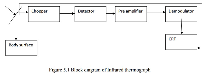
thermo graphic equipment is provided with a special infrared camera that scales
the object. The camera contains an optical system in the form of an oscillating
plane mirror which scans the field of view at a very high speed horizontally
and vertically and focuses the collected infrared radiations onto chopper.
The
chopper disc interrupts the infrared beam so that a.c signals are produced.
Then they are given to detector. The detector is infrared radiation detector.
The detected output by detector is amplified and led to phase sensitive.
2. LIQUID CRYSTAL THERMOGRAPHY
Liquid
crystals are a class of compounds which exhibit colour temperature sensitivity
in the cholestric phase. Scattering effects with the material give rise to iridescent
colours, the dominant wavelength being influenced by very small changes in
temperature.
The high
temperature sensitivity makes cholesteric liquid crystals useful for thermal
mapping.In this technique, the temperature sensitive plate consists of a
blackened thin flim support into which encapsulated liquid crystals cemented to
a pseudo solid powder ( with particle sizes between 10 to 30 ) have been
incorporated.
Thermal
contact between the skin surface and plate produces a color change in the
encapsulated liquid crystals; red for relatively low temperatures through the
visual spectrum to violet for high temperatures. But in infrared thermograms,
the violet colour is used to identify the low temperature regions and the
bright colour or red is used to identify the temperature regions.
If we
want to study a breast’s temperature distribution, several different plates are
necessary to cover a breast temperature range from 280C to360C.
Each plate covers a range of temperature 30C. A record of the liquid
crystal image may be obtained by colour photography. The response time varies
according to the thickness of plate ( ranges from 0.06mm to 0.3 mm) and is 20
to 40 seconds.
3. MICROWAVE THERMOGRAPHY
Eventhough
we get microwave emissions from the skin surface, that intensity is very small
when we compare with Infra red radiation intensity . (10 wavelenght emission
intensity is 108 times greater than 10 cm wavelength emission
intensity). But using modern microwave radiometers one can detect temperature
change of 0.1K. since body tissues are partially transparent to microwave
radiations which orginates from a tissue volume extending from the skin surface
to a depth of several centimeters. Microwave radiometers consisting of matched
antennae placed in contact with the skin surface for use at 1.3 G Hz and 3.3 G
Hz have been used to sense subcutaneous temperature.
The
present day thermographic systems, using Infrared radiation, only give a
temperature map of the skin due to low penetration depth of the short
wavelength of the infrared component of the emitted radiation. Using a
microwave receiver with a frequency response from 1.7 GHz to 2.5 GHz a
penetration depth of 1 cm in tissue and 8 cm in fat and bone can be obtained.
A severe
problem is the unknown emissivity of the body surface for microwaves, as part
of the radiation is reflected back into the body.In a conventional radiometer
this gives rise toa measurement error proportional to the temperature
difference between the body surface and the applied antenna. This error lies in
the order of 1-2 K which is too high for medical applications.
The
problem has been solved iv an elegant way by adding artificial microwave noise
from the antenna,thus providing a radiation balace between the receiver and
body surface. With this a temperature sensitivity of 0.1 K could be obtained.
Based on the transducer attachment on the skin surface, we can classify the
thermography into contact thermography and tele-thermography.
Advantages of Thermography
Get a
visual picture so that you can compare temperatures over a large area It is
real time capable of catching moving targets
Able to
find deteriorating components prior to failure Measurement in areas
inaccessible or hazardous for other methods It is a non-destructive test method
Limitations & disadvantages of thermography
Quality
cameras are expensive and are easily damaged
Images
can be hard to interpret accurately even with experience
Accurate
temperature measurements are very hard to make because of emissivities Most
cameras have ±2% or worse accuracy (not as accurate as contact)
Training
and staying proficient in IR scanning is time consuming Ability to only measure
surface areas
Applications
Healthy
Cases
Tumors
Inflammation
Diseases
of peripheral Vessels
Burns and
Perniones
Skin
Grafts and Organ Transplantation
Collagen
diseases
Orthopedic
Diseases
Brain and
Nervous Diseases
Harmone
Diseases
Examination
of Placenta Attachment
Related Topics