Chapter: Medical Electronics : Recent Trends in Medical Insrumentation
Lasers in Medicine
LASERS IN MEDICINE
LASERS (Light Amplification by Stimulated
Emission of Radiation)
Characteristics
of laser sources
•Tissue
optical properties
•Laser/tissue
interactions
•Some
diagnostic applications
Components of a Laser
a) Lasing
Medium: provides appropriate transition and determines wavelength.Solid: Ruby,
v Nd:YAG, Ti:Sapphire, etc. Liquid: Organic dyes, e.g. rhodamine Gas: Ar, CO2,
HeNe, ArF, etc.
b) Pump:
provides energy necessary for population inversion.
E.g.
electric discharge, flashlamp, another laser.
c) Cavity:
provides opportunity for amplification and produces a directional beam.
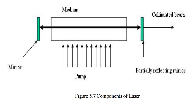
Useful
Characteristics of Output Beam
a) Coherence
b) Collimation
c)
Monochromaticity
d)Widerangeofpulsestructure
e) High
power
Optical Properties of Tissues
Scattering
·
Elastic (i.e. no energy loss), although Doppler
shift and Raman shift have been exploited for diagnostic information.
·
Mean free path for scattering is typically 100
microns.
·
Scattering is forward peaked, typically the average
cosine of the scattering angle is > 0.9 (for isotropic scatt
·
Scattering coefficient decreases slowly as a
function of wavelength.
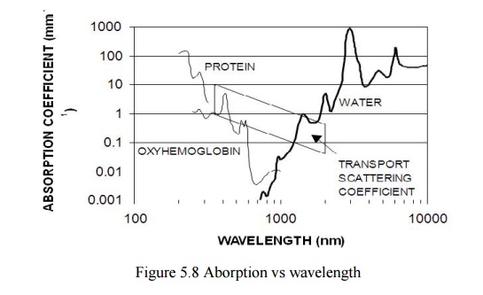
Absorption
Depends
on concentration and absorption spectra of specific molecules in the tissue.
Highly dependent on wavelength. UV - high absorption by proteins.Visible - can
identify specific features of absorption by hemoglobin, melanin, and other
pigments.700 - 900 nm - the “optical window” where tissue absorption is low,
maximum light penetration in tissue.IR - absorption is mainly due to water,
highest at 2.95 microns.
Distribution of Light in Tissue
The
quantity we are usually interested in is the fluence rate. This is defined as
the ratio of total power incident on an infinitesimal sphere to the cross
sectional area of that sphere. The SI unit is W m-2. It is a measure
of how many photons are available per unit volume in the tissue.The fluence
rate distribution in tissue is highly dependent on the absorption and
scattering coefficents of the tissue.
The beam
is incident on tissue at two different wavelengths:300 nm and 700 nm. At 300 nm
the “effective” scattering coefficient is 1 mm-1 and the absorption
coefficient is 10 mm-1. At 700 nm, let us assume the scattering is
the same but the absorption coefficient is only 0.005 mm-1.
Mechanisms of interaction
In order
for light to affect tissue, absorption must take place. The rate at which
energy is deposited in the tissue is given by the product of the fluence rate
(W cm-2) and the linear absorption
coefficient
(cm-1). The rate of energy absorption largely determines whether
photochemical, thermal, or photomechanical effects are dominant.
Photochemical
Initial
absorption by specific molecules.If photon energy is high enough (UV, excimer
laser), direct bond- breaking is possible.Alternatively, the molecule can be
raised to an excited state from which a variety of chemical reactions are
possible such as the generation of free radicals and reactive oxygen species.
Photomechanical
For very
high rates of energy deposition, shock waves can be generated in the tissue by
mechanisms such as bubble expansion/collapse or plasma formation.The mechanical
properties of the tissue govern the propagation of these waves and their
biological effect.
Tissue
can be ablated (i.e. physically removed from the surface, torn or, in the case
of “brittle” tissue, shattered. Interestingly, these two quantities span many
orders of magnitude but their product (the light fluence), varies over a much
smaller range. This emphasizes the point that is is the rate of energy
absorption that determines the nature of the light-tissue interaction.
Selected Applications of Lasers in Medicine
Diagnostic: Goal is to learn something about
the tissue
Therapeutic: Goal is to modify the tissue,
e.g. kill malignant cells.
Optical spectroscopy
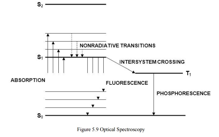
Endogenous absorbers: Hemoglobin,
proteins, melanin, water
Endogenous fluorophores: Collagen, elastin, NADH
Fluorescence Spectroscopy
Noninvasive
tissue characterization to replace or guide physical biopsy, e.g. early
diagnosis of lung cancer.
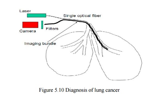
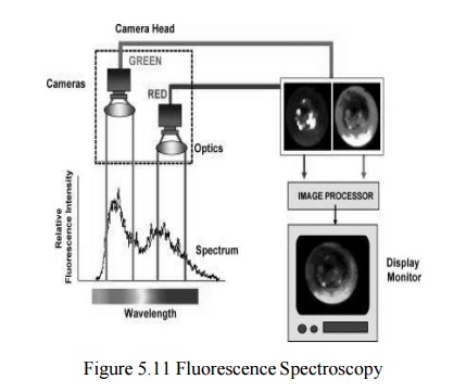
Images are acquired at the two wavelengths shown, and a ratio image is computed and displayed to the physician in real time. This application uses coherence and collimation of the laser toachieve efficient coupling to the fiber and endoscopic light delivery. In addition, the choice of laser (HeCd) provides optical power at the optimum wavelength for fluorescence excitation.
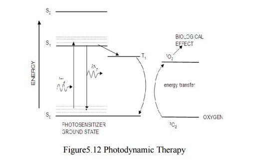
Photodynamic Therapy
Use
chemical reactions initiated by light absorption to kill cells. Original
application in oncology but is applicable to other diseases, including
age-related macular degeneration caused by a proliferation of new blood vessels
in the retina.
Process:
1. Inject photosensitizer or apply topically.
2. Possibly
wait for biodistribution.
3. Irradiate
with light of appropriate wavelength.
Recent advances:
1. Long
wavelength photosensitizers.
2. Reliable
clinical diode lasers.
3. Better
targetting of photosensitizers
Selective Destruction of Blood Vessels
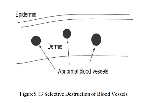
Port Wine Stain: Congenital hypervascularization of
the dermis.Could just ablate the epidermis and dermis but this would result i n
unacceptable scarring. Instead, develop a strategy to target the blood
vessels:
Wavelength;
The vessels are filled with hemoglobin - most of it
oxygenated. Oxyh emoglobin has a strong absorption peak at 577 nm.
Thermal confinement:
Use a pulsed laser to heat the blood in a time
short compared with the “thermal relaxation time of the vessel.
Solution:
Pulsed
dye laser (ms pulses) tuned to 577 nm.
Related Topics