Chapter: Essentials of Anatomy and Physiology: The Nervous System
The Brain - Anatomy and Physiology

THE BRAIN
The brain consists of many parts that function as an integrated whole. The major parts are the medulla, pons, and midbrain (collectively called the brain stem), the cerebellum, the hypothalamus, the thala-mus, and the cerebrum. These parts are shown in Fig. 8–6. We will discuss each part separately, but keep in mind that they are all interconnected and work together.
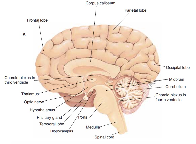
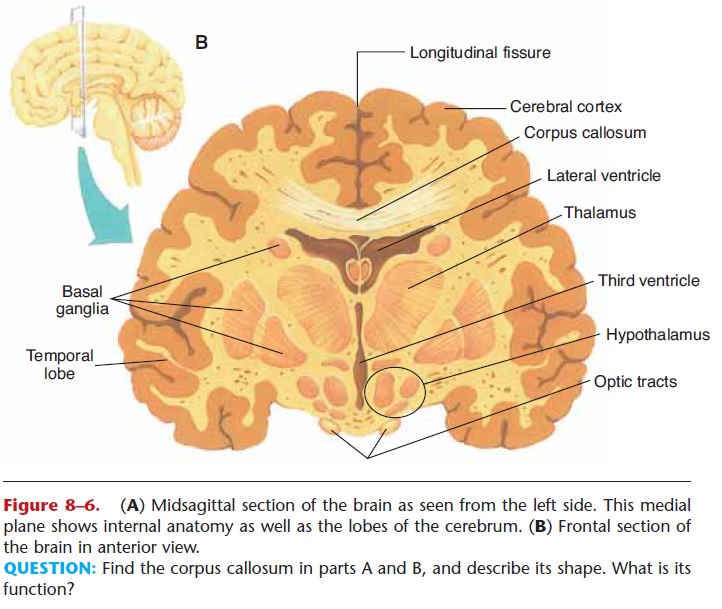
Figure 8–6. (A) Midsagittal section of the brain as seen from the left side. This medial plane shows internal anatomy as well as the lobes of the cerebrum. (B) Frontal section of the brain in anterior view.
QUESTION: Find the corpus callosum in parts A and B, and describe its shape. What is its function?
VENTRICLES
The ventricles are four cavities within the brain: two lateral ventricles, the third ventricle, and the fourth ventricle (Fig. 8–7). Each ventricle contains a capillary network called a choroid plexus, which forms cere-brospinal fluid (CSF) from blood plasma. Cere-brospinal fluid is the tissue fluid of the central nervous system; its circulation and functions will be discussed in the section on meninges.
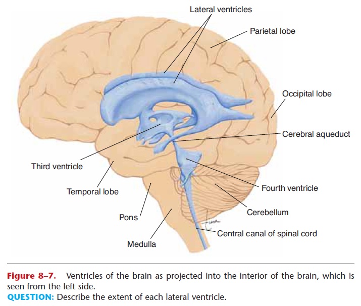
Figure 8–7. Ventricles of the brain as projected into the interior of the brain, which is seen from the left side.
QUESTION: Describe the extent of each lateral ventricle.
MEDULLA
The medulla extends from the spinal cord to the pons and is anterior to the cerebellum. Its functions are those we think of as vital (as in “vital signs”). The medulla contains cardiac centers that regulate heart rate, vasomotor centers that regulate the diameter of blood vessels and, thereby, blood pressure, and respi-ratory centers that regulate breathing. You can see why a crushing injury to the occipital bone may be rapidly fatal—we cannot survive without the medulla. Also in the medulla are reflex centers for coughing, sneezing, swallowing, and vomiting.
PONS
The pons bulges anteriorly from the upper part of the medulla. Within the pons are two respiratory centers that work with those in the medulla to produce a nor-mal breathing rhythm. The many other neurons in the pons ( pons is from the Latin for “bridge”) connect the medulla with other parts of the brain.
MIDBRAIN
The midbrain extends from the pons to the hypothal-amus and encloses the cerebral aqueduct, a tunnel that connects the third and fourth ventricles.
Several different kinds of reflexes are integrated in the mid-brain, including visual and auditory reflexes. If you see a wasp flying toward you, you automatically duck or twist away; this is a visual reflex, as is the coordinated movement of the eyeballs. Turning your head (ear) to a sound is an example of an auditory reflex. The mid-brain is also concerned with what are called righting reflexes, those that keep the head upright and main-tain balance or equilibrium.
CEREBELLUM
The cerebellum is separated from the medulla and pons by the fourth ventricle and is inferior to the occipital lobes of the cerebrum. As you already know, many of the functions of the cerebellum are concerned with movement. These include coordination, regula-tion of muscle tone, the appropriate trajectory and endpoint of movements, and the maintenance of pos-ture and equilibrium. Notice that these are all invol-untary; that is, the cerebellum functions below the level of conscious thought. This is important to permit the conscious brain to work without being overbur-dened. If you decide to pick up a pencil, for example,
the impulses for arm movement come from the cere-brum. The cerebellum then modifies these impulses so that your arm and finger movements are coordinated, and you don’t reach past the pencil.
The cerebellum seems also to be involved in certain sensory functions. For example, if you close your eyes and someone places a tennis ball in one hand and a baseball in the other, could you tell which was which? Certainly you could, by the “feel” of each: the texture and the weight or heft. If you pick up a plastic con-tainer of coffee (with a lid on it) could you tell if the cup is full, half-full, or empty? Again, you certainly could. Do you have to think about it? No. The cere-bellum is, in part, responsible for this ability.
To regulate equilibrium, the cerebellum (and mid-brain) uses information about gravity and movement provided by receptors in the inner ears.
HYPOTHALAMUS
Located superior to the pituitary gland and inferior to the thalamus, the hypothalamus is a small area of the brain with many diverse functions:
1. Production of antidiuretic hormone (ADH) and oxytocin; these hormones are then stored in the posterior pituitary gland. ADH enables the kidneys to reabsorb water back into the blood and thus helps maintain blood volume. Oxytocin causes contractions of the uterus to bring about labor and delivery.
2. Production of releasing hormones (also called releasing factors) that stimulate the secretion of hormones by the anterior pituitary gland. A single example will be given here: The hypothalamus produces growth hormone releasing hormone (GHRH), which stimulates the anterior pituitary gland to secrete growth hormone (GH).
3. Regulation of body temperature by promoting responses such as sweating in a warm environment or shivering in a cold environment.
4. Regulation of food intake; the hypothalamus is believed to respond to changes in blood nutrient levels, to chemicals secreted by fat cells, and to hormones secreted by the gastrointestinal tract. For example, during a meal, after a certain duration of digestion, the small intestine produces a hormone that circulates to the hypothalamus and brings about a sensation of satiety, or fullness, and we tend to stop eating.
5. Integration of the functioning of the autonomic nervous system, which in turn regulates the activity of organs such as the heart, blood vessels, and intestines. This will be discussed in more detail later.
6. Stimulation of visceral responses during emotional situations. When we are angry, heart rate usually increases. Most of us, when embarrassed, will blush, which is vasodilation in the skin of the face. These responses are brought about by the autonomic nervous system when the hypothalamus perceives a change in emotional state. The neurologic basis of our emotions is not well understood, and the visceral responses to emotions are not something most of us can control.
7. Regulation of body rhythms such as secretion of hormones, sleep cycles, changes in mood, or mental alertness. This is often referred to as our biological clock, the rhythms as circadian rhythms, meaning “about a day.” If you have ever had to stay awake for 24 hours, you know how disorienting it can be, until the hypothalamic biological clock has been reset.
THALAMUS
The thalamus is superior to the hypothalamus and inferior to the cerebrum. The third ventricle is a narrow cavity that passes through both the thalamus and hypothalamus. Many of the functions of the thalamus are concerned with sensation. Sensory impulses to the brain (except those for the sense of smell) follow neuron pathways that first enter the thalamus, which groups the impulses before relaying them to the cerebrum, where sensations are felt. For example, holding a cup of hot coffee generates impulses for heat, touch and texture, and the shape of the cup (muscle sense), but we do not experience these as separate sensations.
The thalamus integrates the impulses from the cutaneous receptors and from the cerebellum, that is, puts them together in a sort of electrochemical package, so that the cerebrum feels the whole and is able to interpret the sensation quickly.
Some sensations, especially unpleasant ones such as pain, are believed to be felt by the thalamus. However, the thalamus cannot localize the sensation; that is, it does not know where the painful sensation is. The sensory areas of the cerebrum are required for localization and precise awareness.
The thalamus may also suppress unimportant sensations. If you are reading an enjoyable book, you may not notice someone coming into the room. By temporarily blocking minor sensations, the thalamus permits the cerebrum to concentrate on important tasks.
Parts of the thalamus are also involved in alertness and awareness (being awake and knowing we are), and others contribute to memory. For these functions, as for others, the thalamus works very closely with the cerebrum.
CEREBRUM
The largest part of the human brain is the cerebrum, which consists of two hemispheres separated by longitudinal fissure. At the base of this deep groove is the corpus callosum, a band of 200 million neurons that connects the right and left hemispheres. Within each hemisphere is a lateral ventricle.
The surface of the cerebrum is gray matter called the cerebral cortex. Gray matter consists of cell bodies of neurons, which carry out the many functions of the cerebrum. Internal to the gray matter is white matter, made of myelinated axons and dendrites that connect the lobes of the cerebrum to one another and to all other parts of the brain.
In the human brain the cerebral cortex is folded extensively. The folds are called convolutions or gyri and the grooves between them are fissures or sulci (you can see the folding of the cortex in the frontal section of the brain in Fig. 8–6). This fold-ing permits the presence of millions more neurons in the cerebral cortex. The cerebral cortex of an animal such as a dog or cat does not have this extensive folding. This difference enables us to read, speak, do long division, write poetry and songs, and do so many other “human” things that dogs and cats can-not do.
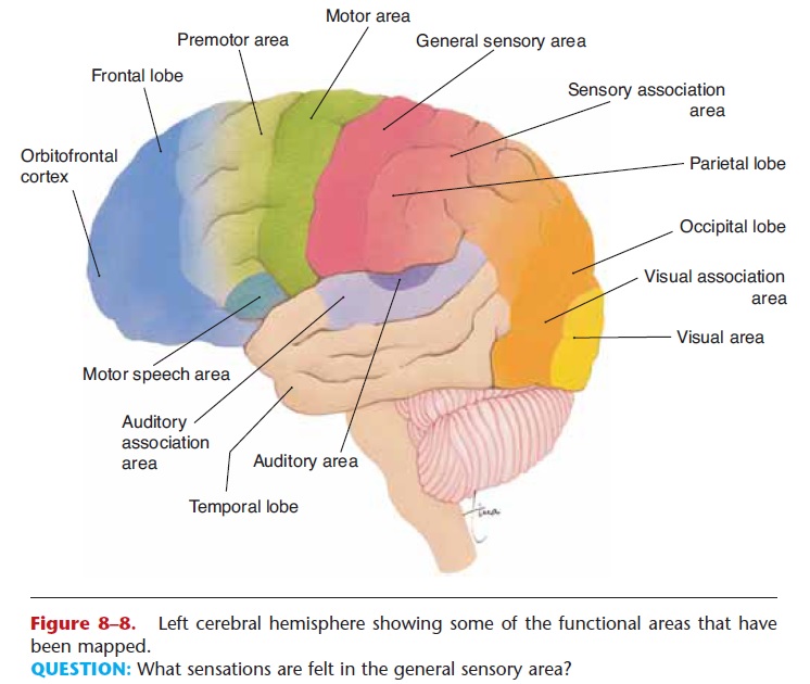
Figure 8–8. Left cerebral hemisphere showing some of the functional areas that have been mapped.
QUESTION: What sensations are felt in the general sensory area?
The cerebral cortex is divided into lobes that have the same names as the cranial bones external to them. Therefore, each hemisphere has a frontal lobe, pari-etal lobe, temporal lobe, and occipital lobe (Fig. 8–8). These lobes have been mapped; that is, certain areas are known to be associated with specific functions. We will discuss the functions of the cerebrum according to these mapped areas.
Frontal Lobes
Within the frontal lobes are the motor areas that generate the impulses for voluntary movement. The largest portions are for movement of the hands and face, those areas with many muscles capable of very fine or precise movements. It is the large size of the motor area devoted to them that gives these muscles their precision. The left motor area controls move-ment on the right side of the body, and the right motor area controls the left side of the body. This is why a patient who has had a cerebrovascular accident, or stroke, in the right frontal lobe will have paralysis of muscles on the left side.
Anterior to the motor areas are the premotor areas, which are concerned with learned motor skills that require a sequence of movements. Tying shoe-laces, for example, seems almost automatic to us; we forget having learned it. It is not a reflex, however; rather the premotor cortex has learned the sequence so well that we are able to repeat it without con-sciously thinking about it.
The parts of the frontal lobes just behind the eyes are the prefrontal or orbitofrontal cortex. This area is concerned with things such as keeping emotional responses appropriate to the situation, realizing that there are standards of behavior (laws or rules of a game or simple courtesy) and following them, and anticipating and planning for the future. An example may be helpful to put all this together: Someone with damage to the prefrontal area might become enraged if his pen ran out of ink during class, might throw the pen at someone, and might not think that a pen will be needed tomorrow and that it is time to go buy one. As you can see, the prefrontal cortex is very important for social behavior, and greatly contributes to what makes us human.
Also in the frontal lobe, usually only the left lobe for most right-handed people, is Broca’s motor speech area, which controls the movements of the mouth involved in speaking.
Parietal Lobes
The general sensory areas in the parietal lobes receive impulses from receptors in the skin and feel and interpret the cutaneous sensations. The left area is for the right side of the body and vice versa. These areas also receive impulses from stretch receptors in muscles for conscious muscle sense. The largest por-tions of these areas are for sensation in the hands and face, those parts of the body with the most cutaneous receptors and the most muscle receptors. The taste areas, which overlap the parietal and temporal lobes, receive impulses from taste buds on the tongue and elsewhere in the oral cavity.
Temporal Lobes
The olfactory areas in the temporal lobes receive impulses from receptors in the nasal cavities for the sense of smell. The olfactory association area learns the meaning of odors such as the smell of sour milk, or fire, or brownies baking in the oven, and enables the thinking cerebrum to use that information effectively.
The auditory areas, as their name suggests, receive impulses from receptors in the inner ear for hearing. The auditory association area is quite large. Part of it is concerned with the meanings of words we hear, that is, with speech. Other parts are for the interpretation of sounds such as thunder during a storm, an ambu-lance siren, or a baby crying. Without proper interpre-tation, we would hear the sound but would not know what it meant, and could not respond appropriately.
Also in the temporal and parietal lobes in the left hemisphere (for most of us) are other speech areas concerned with the thought that precedes speech. Each of us can probably recall (and regret) times when we have “spoken without thinking,” but in actuality that is not possible. The thinking takes place very rap-idly and is essential in order to be able to speak.
Occipital Lobes
Impulses from the retinas of the eyes travel along the optic nerves to the visual areas in the occipital lobes. These areas “see.” The visual association areas inter-pret what is seen, and enable the thinking cerebrum to use the information. Imagine looking at a clock. Seeing the clock is far different from being able to interpret it. At one time we learned to interpret the clock face and hands, and now we do not have to con-sciously decide what time the clock is reading. We can simply use that information, such as hurrying a bit so as not to be late to class. Other parts of the occipital lobes are concerned with spatial relationships; things such as judging distance and seeing in three dimen-sions, or the ability to read a map and relate it to the physical world.
The cerebral cortex has the characteristic of neural plasticity, the ability to adapt to changing needs, to recruit different neurons for certain functions, as may occur during childhood or recovery from a stroke. Another example is the visual cortex of a person who is born blind. The neurons in the occipital lobes that would have been used for vision will often be used for another function; some may become part of an audi-tory area that is used to localize sounds and estimate their distance. Those of us who can see may not rely on hearing for localization; we simply look at where we think the sound came from. A blind person cannot do this, and may have an extensive mental catalogue of sounds, meanings of sounds, distances of sounds, and so on, some of these in the part of the cortex that nor-mally is for vision.
The younger the person, the more plastic the brain. The brains of children are extraordinarily adaptable. As we get older, this ability diminishes, but is still present.
Association Areas
As you can see in Fig. 8–8, many parts of the cerebral cortex are not concerned with movement or a particu- lar sensation. These may be called association areas and perhaps are what truly make us individuals. It is probably these areas that give each of us a personality, a sense of humor, and the ability to reason and use logic. Learning and memory are also functions of these areas.
Although much has been learned about the forma-tion of memories, the processes are still incompletely understood and mostly beyond the scope of this book. Briefly, however, we can say that memories of things such as people or books or what you did last summer involve the hippocampus (from the Greek for “seahorse,” because of its shape), part of the temporal lobe on the floor of the lateral ventricle. The two hip-pocampi seem to collect information from many areas of the cerebral cortex. When you meet a friend, for example, the memory emerges as a whole: “Here’s Fred,” not in pieces. People whose hippocampi are damaged cannot form new memories that last more than a few seconds.
The right hippocampus is also believed to be involved in spatial cognition (literally: “space think-ing”). For example, if you are in school and a friend asks you the shortest way to your home, you will prob-ably quickly form a mental map. You can see how much memory that involves (streets, landmarks, and so on), but the hippocampus can take it a step further and make your memories three-dimensional and men-tally visible. You can see your way home. That is spa-tial cognition.
It is believed that most, if not all, of what we have experienced or learned is stored somewhere in the brain. Sometimes a trigger may bring back memories; a certain scent or a song could act as possible triggers. Then we find ourselves recalling something from the past and wondering where it came from.
The loss of personality due to destruction of brain neurons is perhaps most dramatically seen in Alzheimer’s disease.
Basal Ganglia
The basal ganglia are paired masses of gray matter within the white matter of the cerebral hemispheres (see Fig. 8–6). Their functions are certain subcon-scious aspects of voluntary movement, and they work with the cerebellum. The basal ganglia help regulate muscle tone, and they coordinate accessory move-ments such as swinging the arms when walking or ges-turing while speaking. The most common disorder of the basal ganglia is Parkinson’s disease.
Corpus Callosum
As mentioned previously, the corpus callosum is a band of nerve fibers that connects the left and right cerebral hemispheres. This enables each hemisphere to know of the activity of the other. This is especially important for people because for most of us, the left hemisphere contains speech areas and the right hemi-sphere does not. The corpus callosum, therefore, lets the left hemisphere know what the right hemisphere is thinking about, and the right hemisphere know what the left hemisphere is thinking and talking about. A brief example may be helpful. If you put your left hand behind your back and someone places a pencil in your hand (you are not looking at it) and asks you what it is, would you be able to say? Yes, you would. You would feel the shape and weight of the pencil, find the point and the eraser. The sensory impulses from your left hand are interpreted as “pencil” by the general sensory area in your right parietal lobe. Your right hemisphere probably cannot speak, but its thoughts can be con-veyed by way of the corpus callosum to the left hemi-sphere, which does have speech areas. Your left hemisphere can say that you are holding a pencil. Other aspects of the “division of labor” of our cerebral hemispheres are beyond the scope of this book, but it is a fascinating subject that you may wish to explore further
Related Topics