Chapter: Essentials of Anatomy and Physiology: The Nervous System
Autonomic Nervous System
THE AUTONOMIC NERVOUS SYSTEM
The autonomic nervous system (ANS) is actually part of the peripheral nervous system in that it consists of motor portions of some cranial and spinal nerves. Because its functioning is so specialized, however, the autonomic nervous system is usually discussed as a separate entity, as we will do here.
Making up the autonomic nervous system are vis-ceral motor neurons to smooth muscle, cardiac mus-cle, and glands. These are thevisceral effectors; muscle will either contract or relax, and glands will either increase or decrease their secretions.
The ANS has two divisions: sympathetic and parasympathetic. Often, they function in opposition to each other, as you will see. The activity of both divi-sions is integrated by the hypothalamus, which ensures that the visceral effectors will respond appro-priately to the situation.
AUTONOMIC PATHWAYS
An autonomic nerve pathway from the central nervous system to a visceral effector consists of two motor neurons that synapse in a ganglion outside the CNS (Fig. 8–12). The first neuron is called the pregan-glionic neuron, from the CNS to the ganglion. Thesecond neuron is called the postganglionic neuron, from the ganglion to the visceral effector. The ganglia are actually the cell bodies of the postganglionic neurons.
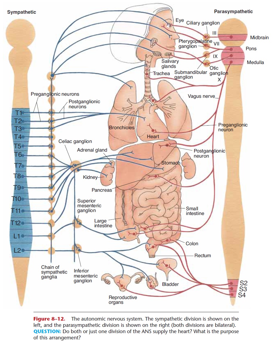
Figure 8–12. The autonomic nervous system. The sympathetic division is shown on the left, and the parasympathetic division is shown on the right (both divisions are bilateral). QUESTION: Do both or just one division of the ANS supply the heart? What is the purpose of this arrangement?
SYMPATHETIC DIVISION
Another name for the sympathetic division is thora-columbar division, which tells us where the sympa-thetic preganglionic neurons originate.
Their cell bodies are in the thoracic segments and some of the lumbar segments of the spinal cord. Their axons extend to the sympathetic ganglia, most of which are located in two chains just outside the spinal column (see Fig. 8–12). Within the ganglia are the synapses between preganglionic and postganglionic neurons; the postganglionic axons then go to the visceral effec-tors. One preganglionic neuron often synapses with many postganglionic neurons to many effectors. This anatomic arrangement has physiological importance: The sympathetic division brings about widespread responses in many organs.
The sympathetic division is dominant in stressful situations, which include anger, fear, or anxiety, as well as exercise. For our prehistoric ancestors, stress-ful situations often involved the need for intense phys-ical activity—the “fight or flight response.” Our nervous systems haven’t changed very much in 50,000 years, and if you look at Table 8–5, you will see the kinds of responses the sympathetic division stimulates. The heart rate increases, vasodilation in skeletal mus-cles supplies them with more oxygen, the bronchioles dilate to take in more air, and the liver changes glyco-gen to glucose to supply energy. At the same time,
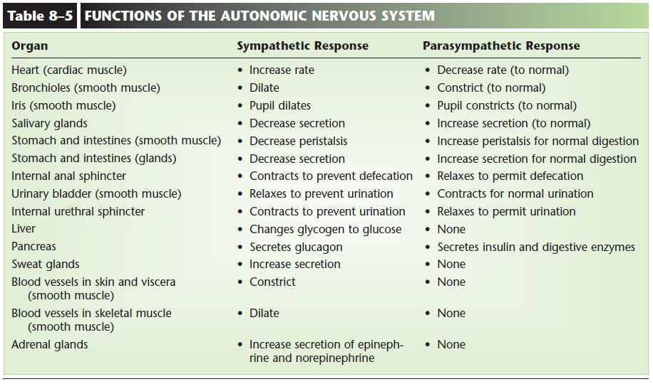
digestive secretions decrease and peristalsis slows; these are not important in a stress situation. Vasoconstriction in the skin and viscera shunts blood to more vital organs such as the heart, muscles, and brain. All of these responses enabled our ancestors to stay and fight or to get away from potential danger. Even though we may not always be in life-threatening situations during stress (such as figuring out our income taxes), our bodies are prepared for just that.
PARASYMPATHETIC DIVISION
The other name for the parasympathetic division is the craniosacral division. The cell bodies of parasym-pathetic preganglionic neurons are in the brain stem and the sacral segments of the spinal cord. Their axons are in cranial nerve pairs 3, 7, 9, and 10 and in some sacral nerves and extend to the parasympathetic gan-glia. These ganglia are very close to or actually in the visceral effector (see Fig. 8–12), and contain the post-ganglionic cell bodies, with very short axons to the cells of the effector.
In the parasympathetic division, one preganglionic neuron synapses with just a few postganglionic neurons to only one effector. With this anatomic arrangement, very localized (one organ) responses are possible.
The parasympathetic division dominates in relaxed (non-stress) situations to promote normal functioning of several organ systems. Digestion will be efficient, with increased secretions and peristalsis; defecation and urination may occur; and the heart will beat at a normal resting rate. Other functions of this division are listed in Table 8–5.
Notice that when an organ receives both sympa-thetic and parasympathetic impulses, the responses are opposites. Such an arrangement makes maintaining an appropriate level of activity quite simple, as in chang-ing the heart rate to meet the needs of a situation. Notice also that some visceral effectors receive only sympathetic impulses. In such cases, the opposite response is brought about by a decrease in sympathetic impulses. Secretion by the sweat glands is an example.
NEUROTRANSMITTERS
Recall that neurotransmitters enable nerve impulses to cross synapses. In autonomic pathways there are two synapses: one between preganglionic and postgan-glionic neurons, and the second between postgan-glionic neurons and visceral effectors.
Acetylcholine is the transmitter released by all preganglionic neurons, both sympathetic and para sympathetic; it is inactivated by cholinesterase in postganglionic neurons. Parasympathetic postgan-glionic neurons all release acetylcholine at the synapses with their visceral effectors. Most sympa-thetic postganglionic neurons release the transmitternorepinephrine at the synapses with the effector cells. Norepinephrine is inactivated by either catechol-O-methyl transferase (COMT) or monoamine oxidase (MAO), or it may be removed from the synapse by reuptake.
Related Topics