Chapter: Human Neuroanatomy(Fundamental and Clinical): The Basal Nuclei
The Basal Nuclei
The Basal Nuclei
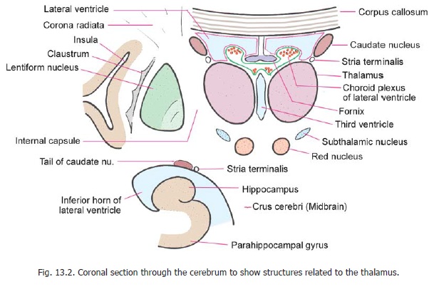
The basal nuclei (or basal ganglia) are large masses of grey matter situated in the cerebral hemispheres. Classically, the following have been included under the definition of basal nuclei (Fig. 13.2). All these are telencephalic in origin.
1. Caudate nucleus.
2. Lentiform nucleus, which consists of two functionally distinct parts, the putamen and the globus pallidus.
3. Amygdaloid nuclear complex.
4. The claustrum is often included among the basal ganglia.
Various other terms commonly used for some of the above nuclei are as follows. The caudate nucleus and the lentiform nucleus together constitute the corpus striatum. This consists of two functionally distinct parts. The caudate nucleus and the putamen form one unit called the striatum, while the globus pallidus forms the other unit, the pallidum.
Recent researches have shown that a number of masses of grey matter, other than those listed above, are very closely related functionally to the basal nuclei. These are as follows.
1. Thesubthalamic nucleus(which is of diencephalic origin) is so closely linked to the basalnuclei that it is now regarded as belonging to this group.
2. Thesubstantia nigra(midbrain) is also closely linked, functionally, to the basal nuclei.
3. Some masses of grey matter found just below the corpus striatum (near the anterior perforatedsubstance) are described as the ventral striatum. The part of the globus pallidus which lies below the level of the anterior commissure is designated as the ventral pallidum.
The Caudate Nucleus
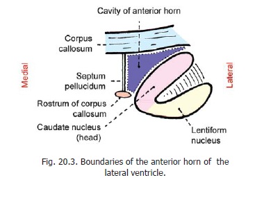
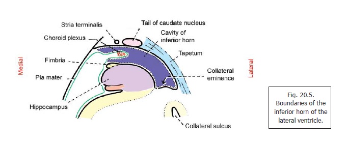
The caudate nucleus is a C-shaped mass of grey matter (Fig.14.1). It consists of a large head, a body and a thin tail. The nucleus is intimately related to the lateral ventricle. The head of the nucleus bulges into the anterior horn of the ventricle and forms the greater part of its floor (Fig.20.3). The body of the nucleus lies in the floor of the central part (Fig. 20.2); and the tail in the roof of the inferior horn of the ventricle (Fig. 20.5). The anterior part of the head of the caudate nucleus is fused, inferiorly, with the lentiform nucleus. This region of fusion is referred to as the fundusstriati. The fundus striati is continuous, inferiorly,with the anterior perforated substance. The anterior end of the tail of the caudate nucleus ends by becoming continuous with the lentiform nucleus. It lies in close relation to the amygdaloid complex.
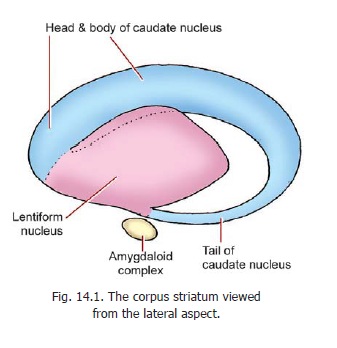
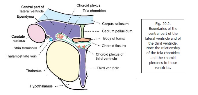
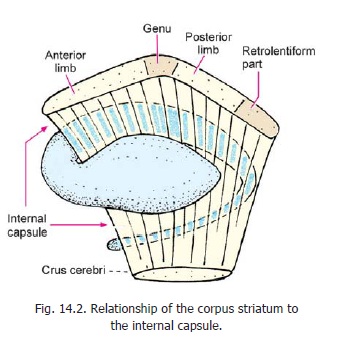
The body of the caudate nucleus is related medially to the thalamus, and laterally to the internal capsule which separates it from the lentiform nucleus (Fig. 14.2). Some other relationships of the caudate nucleus are shown in Fig. 13.2..
The Lentiform Nucleus
The lentiform nucleus lies lateral to the internal capsule. Laterally, it is separated from the claustrum by fibres of the external capsule. (Note that these capsules are so called because they appear, by naked eye, to form a covering for the lentiform nucleus). Superiorly, the lentiform nucleus is related to the corona radiata, and inferiorly to the sublentiform part of the internal capsule. Some other relationships are evident in Fig. 13.2. The lentiform nucleus appears triangular (or wedge shaped) in coronal section. It is divided, by a thin lamina of white matter, into a lateral part, the putamen; and a medial part, the globus pallidus. The globus pallidus is further subdivided into medial and lateral (or internal and external) segments.
The Claustrum
This is a thin lamina of grey matter that lies lateral to the lentiform nucleus. It is separated from the latter by fibres of the external capsule. Laterally, it is separated by a thin layer of white matter from the cortex of the insula. Its connections and functions are unknown.
The Amygdaloid Complex
This complex (also called the amygdaloid body, amygdala) lies in the temporal lobe of the cerebral hemisphere, close to the temporal pole. It lies deep to the uncus, and is related to the anterior end of the inferior horn of the lateral ventricle.
Connections of the Basal Nuclei
The various basal nuclei have numerous connections, but no useful purpose is served by enumerating the afferents and efferents of each nucleus. An integrated view of the corpus striatum, along with the substantia nigra and the subthalamic nucleus, is necessary. A scheme showing the main connections is given in Fig. 14.3.
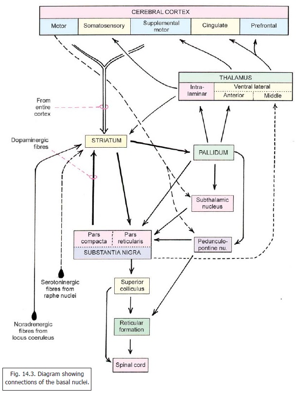
The striatum (caudate nucleus and putamen) receive afferents from the following.
1. The entire cerebral cortex. These fibres are glutamatergic.
2. The intralaminar nuclei of the thalamus.
3. The pars compacta of the substantia nigra.
These fibres are dopaminergic. (Some dopaminergic fibres arise from the retrorubral nuclei lying behind the red nucleus).
4. Noradrenergic fibres are received from the raphe nuclei (in the reticular formation of the midbrain).
5. Serotoninergic fibres are received from the locus coeruleus.
The afferents from the cerebral cortex and from the thalamus provide the striatum with various modalities of sensory information (other than olfactory). The main output of the striatum is concentrated upon the pallidum, and on the substantia nigra (pars reticularis). Fibres also reach the substantia nigra from the pallidum directly, or after relay in the subthalamic nucleus or in the pedunculo-pontine nucleus.
The efferents of the pallidum are as follows.
1. Like the striatum, the pallidum projects to the substantia nigra. These fibres take three mainroutes. Some reach the substantia nigra directly. Others are relayed in the subthalamic nucleus; while still others are relayed in the pedunculopontine nucleus,
2. The pallidum projects to theintralaminar nuclei of the thalamus, from where impulses are relayed to the somatosensory area of the cerebral cortex.
3. The pallidum also projects to theanterior part of the ventral lateral nucleus of the thalamus. These inputs are relayed to the supplemental motor area.
Connections of substantia nigra
As shown in Fig. 14.3 the pars compacta of the substantia nigra sends a dopaminergic projection to the striatum. A projection from the striatum ends in the pars reticularis of the substantia nigra. This part also receives fibres from the pallidum directly, or after relay in the subthalamic nucleus or in the pedunculo-pontine nucleus.
The pars reticularis projects to the (middle part of the) ventral lateral nucleus of the thalamus. These impulses are relayed to cingulate and prefrontal areas of the cerebral cortex. Other efferents of the pars reticularis reach the superior colliculus. They are relayed from there to the reticular formation of the medulla, and to the spinal cord. These regions also receive fibres descending from the pedunculopontine nucleus.
In summary note the following.
a. The various basal nuclei are interconnected. The substantia nigra, the subthalamic nucleus, and the pedunculo-pontine nucleus form an integral part of this interconnected system.
b. Cranially, the system receives fibres from the cerebral cortex; and send back impulses to it through the thalamus.
c. Descending fibres from the system influence the superior colliculus, the reticular formation of the medulla, and the spinal cord.
Ventral striatum and pallidum
On the basis of recent investigations some masses of grey matter lying in the region of the anterior perforated substance are now described as the ventral striatum. In Fig. 14.4 we see the anterior part of the caudate and lentiform nuclei. Inferiorly, the two nuclei fuse to the form the fundus striati.
Immediately below the fundus striati we see the olfactory tubercle (in the anterior perforated substance). More medially, we see a mass of grey matter called the nucleus accumbens. Note that this nucleus is closely related to the caudate nucleus (superolaterally) and to the septal nuclei medially. The ventral striatum consists of the nucleus accumbens and the olfactory tubercle.
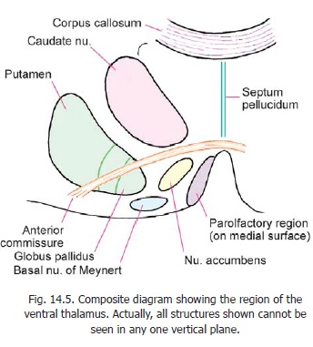
A coronal section through the brain a little posterior to the plane of Fig. 14.4 is shown in Fig. 14.5. Note the anterior commissure running laterally just below the head of the caudate nucleus. It cuts through the globus pallidus. The part of the globus pallidus lying inferior to the anterior commissure is called the ventral pallidum. Identify the olfactory tubercle in Fig. 14.5. Medial to it there is a collection of neurons that form the basal nucleus of Meynert. In this figure the position of the nucleus accumbens is shown diagrammatically: it actually lies more anteriorly.
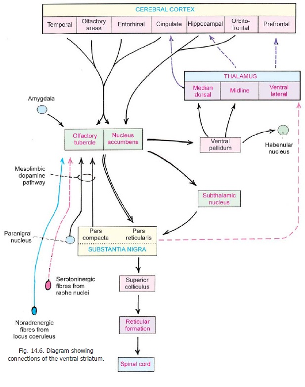
The reason for considering the nucleus accumbens and the olfactory tubercle as parts of the striatum is that their connections are very similar to those of the main part of the striatum (or dorsal striatum). These are shown in Fig. 14.6 which should be compared with Fig. 14.3.
Related Topics