Chapter: Essentials of Anatomy and Physiology: The Integumentary System
Subcutaneous Tissue
SUBCUTANEOUS TISSUE
The subcutaneous tissue may also be called the superficial fascia, one of the connective tissue membranes. Made of areolar connective tissue and adipose tissue, the superficial fascia connects the dermis to the underlying muscles.
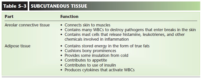
Areolar connective tissue, or loose connective tis-sue, contains collagen and elastin fibers and many white blood cells that have left capillaries to wander around in the tissue fluid between skin and muscles. These migrating white blood cells destroy pathogens that enter the body through breaks in the skin. Mast cells are specialized connective tissue cells found in areolar tissue; they produce histamine, leukotrienes, and other chemicals that help bring about inflammation, the body’s response to damage.
The cells (adipocytes) of adipose tissue are specialized to store fat, and our subcutaneous layer of fat stores excess nutrients as a potential energy source. This layer also cushions bony prominences, such as when sitting, and provides some insulation from cold. For people, this last function is relatively minor, because we do not have a thick layer of fat, as do ani-mals such as whales and seals. As mentioned, adipose tissue is involved in the onset or cessation of eating and in the use of insulin by body cells, and it contributes to inflammation by produc-ing cytokines, chemicals that activate white blood cells.
Just as the epidermis forms a continuous sheet that covers the body, the subcutaneous tissue is a continu-ous layer, though it is internal. If we consider the epi-dermis as the first line of defense against pathogens, we can consider the subcutaneous tissue a secondary line of defense. There is, however, a significant anatomic difference. The cells of the epidermis are very closely and tightly packed, but the cells and protein fibers of subcutaneous tissue are farther apart, and there is much more tissue fluid. If we imagine the epidermis as a four-lane highway during rush hour with bumper-to-bumper traffic, the superficial fascia would be that same highway at three o’clock in the morning, when it is not crowded and cars can move much faster. This is an obvious benefit for the migrat-ing white blood cells, but may become a disadvantage because some bacterial pathogens, once established, can spread even more rapidly throughout subcuta-neous tissue.
Group A streptococcus, for example, is a cause of necrotizing fasciitis. Necrotizing means “to cause death,” and fasciitis is the inflammation of a fascia, in this case the superficial fascia and the deep fasciae around muscles. Necrotizing fasciitis is an extremely serious infection and requires surgical removal of the infected tissue, or even amputation of an infected limb, to try to stop the spread of the bacteria. The body’s defenses have been overwhelmed because what is usually an anatomic benefit, the “looseness” of are-olar connective tissue, has been turned against us and become a detriment.
The functions of subcutaneous tissue are summa-rized in Table 5–3.
Related Topics