Chapter: Essentials of Anatomy and Physiology: The Integumentary System
Skin - Anatomy and Physiology
THE SKIN
The two major layers of the skin are the outer epidermis and the inner dermis. Each of these layers is made of different tissues and has very different func-tions.
EPIDERMIS
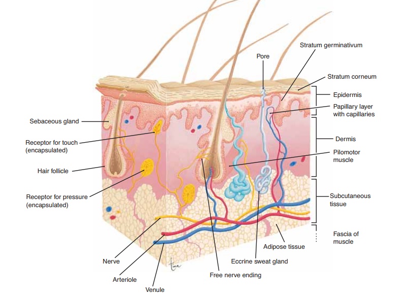
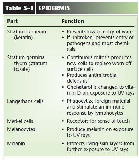
The epidermis is made of stratified squamous kera-tinizing epithelial tissue and is thickest on the palms and soles. The cells that are most abundant are called keratinocytes, and there are no capillaries present between them. Although the epidermis may be further subdivided into four or five sublayers, two of these are of greatest importance: the innermost layer, the stra-tum germinativum, and the outermost layer, the stra-tum corneum (Fig. 5–1).
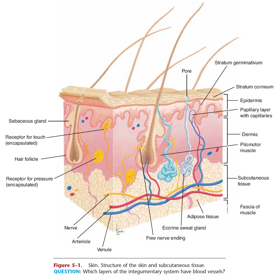
Figure 5–1. Skin. Structure of the skin and subcutaneous tissue.
QUESTION: Which layers of the integumentary system have blood vessels?
Stratum Germinativum
The stratum germinativum may also be called the stratum basale. Each name tells us something about this layer. To germinate means “to sprout” or “to grow.” Basal means the “base” or “lowest part.” The stratum germinativum is the base of the epidermis, the innermost layer in which mitosis takes place. New cells are continually being produced, pushing the older cells toward the skin surface. These cells pro-duce the protein keratin, and as they get farther away from the capillaries in the dermis, they die. As dead cells are worn off the skin’s surface, they are replaced by cells from the lower layers. Scattered among the keratinocytes of the stratum germinativum are very different cells called Merkel cells (or Merkel discs); these are receptors for the sense of touch (Fig. 5–2).
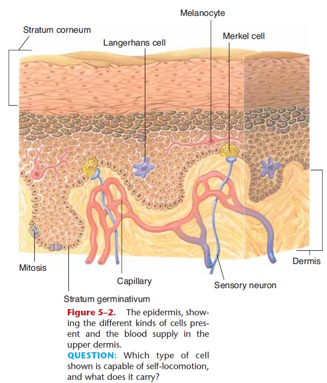
Figure 5–2. The epidermis, show-ing the different kinds of cells pres-ent and the blood supply in the upper dermis.
QUESTION: Which type of cell shown is capable of self-locomotion, and what does it carry?
The living keratinocytes are able to synthesize antimicrobial peptides called defensins; these and other chemicals are produced following any injury to the skin, as part of the process of inflammation. Defensins rupture the membranes of pathogens such as bacteria that may enter by way of breaks in the skin.
The living portion of the epidermis also produces a vitamin; the cells have a form of cholesterol that, on exposure to ultraviolet light, is changed to vitamin D (then modified by the liver and kidneys to the most active form, called 1,25-D, or calcitriol, which is con-sidered a hormone). This is why vitamin D is some-times referred to as the “sunshine vitamin.” People who do not get much sunlight depend more on nutri-tional sources of vitamin D, such as fortified milk. But sunlight is probably the best way to get vitamin D, and 15 minutes a day a few times a week is often enough. Vitamin D is important for the absorption of calcium and phosphorus from food in the small intestine; this function has been known for years. Recent research, however, suggests that vitamin D is also involved in maintaining muscle strength, especially in elderly peo-ple, in the functioning of insulin, and in some immune responses, where it may be protective for some types of cancer.
Stratum Corneum
The stratum corneum, the outermost epidermal layer, consists of many layers of dead cells; all that is left is their keratin. The protein keratin is relatively waterproof, and though the stratum corneum should not be thought of as a plastic bag encasing the body, it does prevent most evaporation of body water. Also of importance, keratin prevents the entry of water. With-out a waterproof stratum corneum, it would be impos-sible to swim in a pool or even take a shower without damaging our cells.
The stratum corneum is also a barrier to pathogens and chemicals. Most bacteria and other microorgan-isms cannot penetrate unbroken skin. The flaking of dead cells from the skin surface helps remove micro-organisms, and the fatty acids in sebum help inhibit their growth. Most chemicals, unless they are corro-sive, will not get through unbroken skin to the living tissue within. One painful exception is the sap of poi-son ivy. This resin does penetrate the skin and initiates an allergic reaction in susceptible people. The inflam-matory response that characterizes allergies causes blisters and severe itching. The importance of the stratum corneum becomes especially apparent when it is lost.
Certain minor changes in the epidermis are undoubtedly familiar to you. When first wearing new shoes, for example, the skin of the foot may be sub-jected to friction. This will separate layers of the epi- dermis, or separate the epidermis from the dermis, and tissue fluid may collect, causing a blister. If the skin is subjected to pressure, the rate of mitosis in the stratum germinativum will increase and create a thicker epidermis; we call this a callus. Although cal-luses are more common on the palms and soles, they may occur on any part of the skin.
Langerhans Cells
Within the epidermis are Langerhans cells, which are also called dendritic cells because of their branched appearance when they move (see Fig. 5–2). These cells originate in the red bone marrow, and are quite mobile. They are able to phagocytize foreign material, such as bacteria that enter the body through breaks in the skin. With such ingested pathogens, the Langerhans cells migrate to lymph nodes and present the pathogen to lymphocytes, a type of white blood cell. This triggers an immune response such as the production of antibodies (antibodies are proteins that label foreign material for destruction). Because of its position adjacent to the external environment, the skin is an important component of the body’s protective responses, though many of the exact aspects of this have yet to be determined.
Melanocytes
Another type of cell found in the lower epidermis is the melanocyte, which is also shown in Fig. 5–2.
melanin. People of the same size have approxi-mately the same number of melanocytes, though these cells may differ in their level of activity. In people with dark skin, the melanocytes continuously produce large amounts of melanin. The melanocytes of light-skinned people produce less melanin. The activity of melanocytes is genetically regulated; skin color is one of our hereditary characteristics.
In all people, melanin production is increased by exposure of the skin to ultraviolet rays, which are part of sunlight and are damaging to living cells. As more melanin is produced, it is taken in by the epidermal cells as they are pushed toward the surface. This gives the skin a darker color, which prevents further expo-sure of the living stratum germinativum to ultraviolet rays. People with dark skin already have good protec-tion against the damaging effects of ultraviolet rays; people with light skin do not. Melanin also gives color to hair, though its protective function is confined to the hair of the head. Two parts of the eye obtain their color from melanin: the iris and the interior choroid layer of the eyeball.The functions of the epidermis and its cells are summarized in Table 5–1.
DERMIS
The dermis is made of an irregular type of fibrous connective tissue, irregular meaning that the fibers are not parallel, but run in all directions. Fibroblasts pro-duce both collagen and elastin fibers. Recall that col-lagen fibers are strong, and elastin fibers are able to recoil after being stretched. Strength and elasticity are two characteristics of the dermis. With increasing age, however, the deterioration of the elastin fibers causes the skin to lose its elasticity. We can all look forward to at least a few wrinkles as we get older.
The uneven junction of the dermis with the epider-mis is called the papillary layer (see Fig. 5–1). Capil-laries are abundant here to nourish not only the dermis but also the stratum germinativum. The epi-dermis has no capillaries of its own, and the lower, liv-ing cells depend on the blood supply in the dermis for oxygen and nutrients.
Within the dermis are the accessory skin structures: hair and nail follicles, sensory receptors, and several types of glands. Some of these project through the epidermis to the skin surface, but their active portions are in the dermis.
HairFollicles
Hair follicles are made of epidermal tissue, and the growth process of hair is very similar to growth of the epidermis. At the base of a follicle is the hair root, which contains cells called the matrix, where mitosis takes place (Fig. 5–3). The new cells produce keratin, get their color from melanin, then die and become incorporated into the hair shaft, which is pushed toward the surface of the skin. The hair that we comb and brush every day consists of dead, keratinized cells. The rate of hair growth averages 0.3 to 0.4 in./month (8 to 10 mm).
Compared to some other mammals, humans do not have very much hair. The actual functions of human hair are quite few. Eyelashes and eyebrows help to keep dust and perspiration out of the eyes, and the hairs just inside the nostrils help to keep dust out of the nasal cavities. Hair of the scalp does provide insu-lation from cold for the head. The hair on our bodies, however, no longer serves this function, but we have the evolutionary remnants of it. Attached to each hair follicle is a small, smooth muscle called the pilomotor
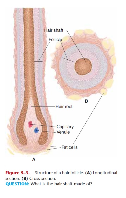
Figure 5–3. Structure of a hair follicle. (A) Longitudinal section. (B) Cross-section.
QUESTION: What is the hair shaft made of?
or arrector pili muscle. When stimulated by cold or emotions such as fear, these muscles pull the hair fol-licles upright. For an animal with fur, this would trap air and provide greater insulation. Because people do not have thick fur, all this does for us is give us “goose bumps.”
Nail Follicles
Found on the ends of fingers and toes, nail follicles produce nails just as hair follicles produce hair. Mitosis takes place in the nail root at the base of the nail (Fig. 5–4), and the new cells produce keratin (a stronger form of this protein than is found in hair) and then die. Although the nail itself consists of keratinized dead cells, the flat nail bed is living epidermis and dermis. This is why cutting a nail too short can be quite painful. Nails protect the ends of the fingers and toes from mechanical injury and give the fingers greater ability to pick up small objects. Fingernails are also good for scratching. This is more important than it may seem at first. An itch might mean the presence of an arthropod parasite, mosquito, tick, flea, or louse. These parasites (all but the tick are insects) feed on blood, and all are potential vectors of diseases caused by bacteria, viruses, or protozoa. A quick and vigorous scratch may kill or at least dislodge the arthropod and prevent the transmission of the disease. Fingernails grow at the rate of about 0.12 in./month (3 mm), and growth is a little faster during the summer months.
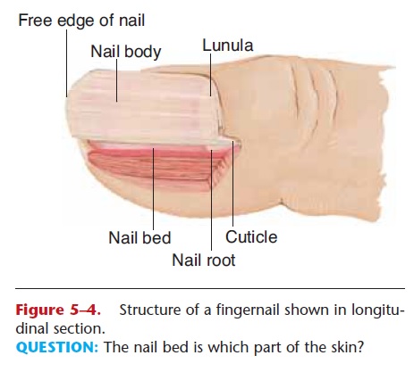
Figure 5–4. Structure of a fingernail shown in longitu-dinal section.
QUESTION: The nail bed is which part of the skin?
Receptors
Most sensory receptors for the cutaneous senses are found in the dermis (Merkel cells are in the stra-tum germinativum, as are some nerve endings). The cutaneous senses are touch, pressure, heat, cold, and pain. For each sensation there is a specific type of receptor, which is a structure that will detect a partic-ular change. For pain, heat, and cold, the receptors are free nerve endings. For touch and pressure, the receptors are called encapsulated nerve endings, which means there is a cellular structure around the sensory nerve ending (see Fig. 5–1). The purpose of these receptors and sensations is to provide the central nervous system with information about the external environment and its effect on the skin. This informa-tion may stimulate responses, such as washing a painful cut finger, scratching an insect bite, or responding to a feeling of cold by putting on a sweater.
The sensitivity of an area of skin is determined by how many receptors are present. The skin of the fin-gertips, for example, is very sensitive to touch because there are many receptors per square inch. The skin of the upper arm, with few touch receptors per square inch, is less sensitive.
When receptors detect changes, they generate nerve impulses that are carried to the brain, which interprets the impulses as a particular sensation. Sensation, therefore, is actually a function of the brain.
Glands
Glands are made of epithelial tissue. The exocrine glands of the skin have their secretory portions in the dermis. Some of these are shown in Fig. 5–1.
Sebaceous Glands. The ducts of sebaceous glands open into hair follicles or directly to the skin surface. Their secretion is sebum, a lipid substance that we commonly refer to as oil. As mentioned previously, sebum inhibits the growth of bacteria on the skin sur-face. Another function of sebum is to prevent drying of skin and hair. The importance of this may not be read-ily apparent, but skin that is dry tends to crack more easily. Even very small breaks in the skin are potential entryways for bacteria. Decreased sebum production is another consequence of getting older, and elderly peo-ple often have dry and more fragile skin.
Adolescents may have the problem of overactive sebaceous glands. Too much sebum may trap bacteria within hair follicles and create small infections. Because sebaceous glands are more numerous around the nose and mouth, these are common sites of pim-ples in young people.
Ceruminous Glands. These glands are located in the dermis of the ear canals. Their secretion is called cerumen or ear wax (which includes the sebum secreted in the ear canals). Cerumen keeps the outer surface of the eardrum pliable and prevents drying.
However, if excess cerumen accumulates in the ear canal, it may become impacted against the eardrum. This might diminish the acuity of hearing by prevent-ing the eardrum from vibrating properly.
Sweat Glands. There are two types of sweat glands, apocrine and eccrine. Apocrine glands are most numerous in the axillae (underarm) and genital areas and are most active in stressful and emotional situa-tions. Although their secretion does have an odor, it is barely perceptible to other people. Animals such as dogs, however, can easily distinguish among people because of their individual scents. If the apocrine secretions are allowed to accumulate on the skin, bac-teria metabolize the chemicals in the sweat and pro-duce waste products that have distinct odors that many people find unpleasant.
Eccrine glands are found all over the body but are especially numerous on the forehead, upper lip, palms, and soles. The secretory portion of these glands is simply a coiled tube in the dermis. The duct of this tube extends to the skin’s surface, where it opens into apore.
The sweat produced by eccrine glands is important in the maintenance of normal body temperature. In a warm environment, or during exercise, more sweat is secreted onto the skin surface, where it is then evapo-rated by excess body heat. Recall that water has a high heat of vaporization, which means that a great deal of heat can be lost in the evaporation of a relatively small amount of water. Although this is a very effective mechanism of heat loss, it has a potentially serious dis-advantage. Loss of too much body water in sweat may lead to dehydration, as in heat exhaustion or even after exercise on a hot and humid day. Increased sweating during exercise or on warm days should always be accompanied by increased fluid intake.
Those who exercise regularly know that they must replace salt as well as water. Sodium chloride is also lost in sweat, as are small amounts of urea (a nitroge-nous waste product of amino acid metabolism). This excretory function of the skin is very minor, however; the kidneys are primarily responsible for removing waste products from the blood and for maintaining the body’s proper salt-to-water proportion.
Blood Vessels
Besides the capillaries in the dermis, the other blood vessels of great importance are the arterioles. Arterioles are small arteries, and the smooth muscle
However, if excess cerumen accumulates in the ear canal, it may become impacted against the eardrum. This might diminish the acuity of hearing by prevent-ing the eardrum from vibrating properly.
Sweat Glands. There are two types of sweat glands, apocrine and eccrine. Apocrine glands are most numerous in the axillae (underarm) and genital areas and are most active in stressful and emotional situa-tions. Although their secretion does have an odor, it is barely perceptible to other people. Animals such as dogs, however, can easily distinguish among people because of their individual scents. If the apocrine secretions are allowed to accumulate on the skin, bac-teria metabolize the chemicals in the sweat and pro-duce waste products that have distinct odors that many people find unpleasant.
Eccrine glands are found all over the body but are especially numerous on the forehead, upper lip, palms, and soles. The secretory portion of these glands is simply a coiled tube in the dermis. The duct of this tube extends to the skin’s surface, where it opens into apore.
The sweat produced by eccrine glands is important in the maintenance of normal body temperature. In a warm environment, or during exercise, more sweat is secreted onto the skin surface, where it is then evapo-rated by excess body heat. Recall that water has a high heat of vaporization, which means that a great deal of heat can be lost in the evaporation of a relatively small amount of water. Although this is a very effective mechanism of heat loss, it has a potentially serious dis-advantage. Loss of too much body water in sweat may lead to dehydration, as in heat exhaustion or even after exercise on a hot and humid day. Increased sweating during exercise or on warm days should always be accompanied by increased fluid intake.
Those who exercise regularly know that they must replace salt as well as water. Sodium chloride is also lost in sweat, as are small amounts of urea (a nitroge-nous waste product of amino acid metabolism). This excretory function of the skin is very minor, however; the kidneys are primarily responsible for removing waste products from the blood and for maintaining the body’s proper salt-to-water proportion.
Blood Vessels
Besides the capillaries in the dermis, the other blood vessels of great importance are the arterioles. Arterioles are small arteries, and the smooth muscle in their walls permits them to constrict (close) or dilate (open). This is important in the maintenance of body temperature, because blood carries heat, which is a form of energy.
In a warm environment the arterioles dilate (vasodilation), which increases blood flow through the dermis and brings excess heat close to the body surface to be radiated to the environment. In a cold environ-ment, however, body heat must be conserved if possi-ble, so the arterioles constrict. The vasoconstriction decreases the flow of blood through the dermis and keeps heat within the core of the body. This adjusting mechanism is essential for maintaining homeostasis. Regulation of the diameter of the arterioles in response to external temperature changes is controlled by the nervous system. These changes can often be seen in light-skinned people. Flushing, especially in the face, may be observed in hot weather. In cold, the skin of the extremities may become even paler as blood flow through the dermis decreases. In people with dark skin, such changes are not as readily apparent because they are masked by melanin in the epidermis.
Vasoconstriction in the dermis may also occur dur-ing stressful situations. For our ancestors, stress usu-ally demanded a physical response: Either stand and fight or run away to safety. This is called the “fight or flight response.” Our nervous systems are still pro-grammed to respond as if physical activity were neces-sary to cope with the stress situation. Vasoconstriction in the dermis will shunt, or redirect, blood to more vital organs such as the muscles, heart, and brain. In times of stress, the skin is a relatively unimportant organ and can function temporarily with a minimal blood flow. You have probably heard the expression “broke out in a cold sweat,” and may even have felt it in a stressful situation. Such sweating feels cold because vasoconstriction in the dermis makes the skin relatively cool.
Blood flow in the dermis may be interrupted by prolonged pressure on the skin. For example, a hospi-tal patient who cannot turn over by herself may develop a decubitus ulcer, also called a pressure ulcer or pressure sore. The skin is compressed between the object outside, such as a bed, and a bony prominence within, such as the heel bone or the sacrum in the lower back. Without its blood supply the skin dies, and the dead tissue is a potential site for bacterial infection.
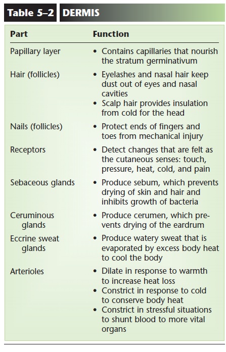
The functions of dermal structures are summarized in Table 5–2.
Related Topics