Chapter: 10th Science : Chapter 14 : Transportation in Plants and Circulation in Animals
Structure of Human Heart
Structure
of Human Heart
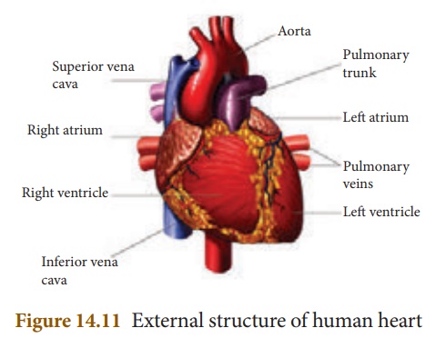
Heart is a muscular
pumping organ that pumps out the blood into the blood vessels. Human heart is
situated between the lungs, slightly tilted toward the left and above the
diaphragm in the thoracic cavity. The heart is made of specialized type
of muscle called the cardiac muscle.
The heart is enclosed in
a double walled sac called pericardium. It contains lubricating pericardial
fluid which reduces friction during heart beat and protects
it from mechanical injuries.![]()
![]()
The human heart is four
chambered. The two upper thin walled chambers of the heart are called
auricle or atria (sing: atrium) and two lower thick walled
chambers are called ventricles. The chambers are separated by partition
called septum. The septum between auricles and ventricles prevents the
mixing of oxygenated and deoxygenated blood.
The two auricles are
separated from each other by interatrial septum.The left atrium is
smaller than the right atrium. The right atrium receives deoxygenated blood
from different parts of the body through the main veins superior vena
cava, inferior vena cava and coronary sinus. Pulmonary veins bring
oxygenated blood to the left atrium from the lungs. The right and
left auricles pump blood into the right and left ventricles respectively.
The ventricles form the
lower part of the heart. The two ventricles are separated from each other by an
interventricular septum. The left and right ventricles have thick
walls because the ventricles have to pump out blood with force away from
the heart. From the right ventricle arises the pulmonary trunk
which bifurcates to form right and left pulmonary arteries. The right and left
pulmonary arteries supply deoxygenated blood to the lungs of the
respective side. The left ventricle is longer and narrower than the
right ventricle. The walls are about three times thicker than the right
ventricle. The left ventricle gives rise to aorta. The oxygenated
blood is supplied by the aorta to various organs of the body. The coronary
arteries supply blood to the heart.
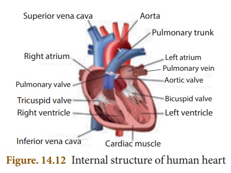
Valves: The valves are the
muscular flaps that regulate the flow of blood in a single direction and
prevent back flow of blood. The heart contains three types of valves.
Right atrioventricular
valve: It
is located between the right auricle and right ventricle. It has three
thin triangular leaf like flaps and therefore called tricuspid valve.
The apices of the flaps are held in position by chordae tendinae arising
from the muscular projection of the ventricle wall known as papillary
muscles.
Left atrioventricular valve: It is located between the left auricle and left ventricle. It has two cusps and therefore called bicuspid or mitral valve.
Semilunar valves: The major arteries (pulmonary
artery and aorta) which leave the heart have semilunar valves which prevent
backward flow of blood into the ventricles. They are the pulmonary and aortic
semilunar valves.
![]()
![]()
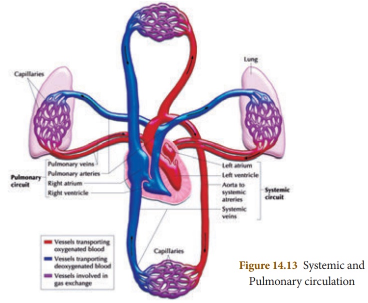
1. Types of Blood Circulation
The blood circulates in
our body as oxygenated and deoxygenated blood. The types of circulation are:
i. Systemic circulation:
Circulation of
oxygenated blood from the left ventricle of the heart to various organs of the
body and return of deoxygenated blood to the right atrium. Aorta carries
oxygenated blood to all the organs of the body.
ii. Pulmonary
circulation: The path of pulmonary circulation starts in the right
ventricle. Pulmonary artery arises from the right ventricle and reaches the
lungs with deoxygenated blood. Pulmonary veins collect the oxygenated blood
from the lungs and supplies it to the left atrium of the heart.
iii. Coronary
circulation: The supply of blood to the heart muscles (cardiac muscles)
is called as coronary circulation. Cardiac muscles receive oxygenated
blood from coronary arteries that originate from the aortic
arch. Deoxygenated blood from the cardiac muscles drains into the
right atrium by the coronary sinuses.
When the blood
circulates twice through the heart in one complete cycle it is called double
circulation. In double circulation the oxygenated blood do not mix
with the deoxygenated blood.
However, in some animals
the oxygenated and deoxygenated blood are mixed and pass through the heart only
once. This type of circulation is called single circulation. e.g.,
fishes, amphibians and certain reptiles.
2. Heart Beat
One complete contraction
(systole) and relaxation (diastole) of the atrium and
ventricles of the heart constitute heartbeat. The heart normally beats 72 – 75
times per minute.
Initiation and conduction of Heart beat
The human heart is myogenic
in nature. Contraction is initiated by a specialized portion of the heart
muscle, the sino-atrial (SA) node which is situated in the wall
of the right atrium near the opening of the superior vena cava. The SA
node is broader at the top and tapering below. It is made up of thin fibres.
Sino-atrial node acts as the
‘pacemaker’ of the heart because it is capable of initiating impulse
which can stimulate the heart muscles to contract. The impulse from the
sinoatrial node spreads as a wave of contraction over the right and left atrial
wall pushing the blood through the atrioventricular valves into the
ventricles. The wave of contraction from SA node reaches the atrioventricular
(AV) node which is stimulated to emit an impulse of contraction
spreading to the ventricular muscle via the atrioventricular bundle and the
Purkinje fibres.
Pulse: When the heart beats the
blood is forced into the arteries. The expansion of the artery every
time the blood is forced into it is called pulse. It can be felt by placing the
fingertip on the artery near the wrist. Normal pulse rate ranges from 70 – 90 /
min.
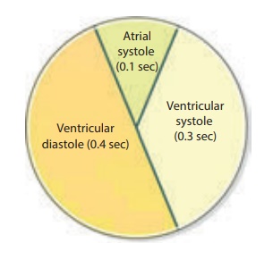
3. Cardiac Cycle
The sequence of events
occurring from the beginning to the completion of one heart beat
is called cardiac cycle. During cardiac cycle blood flows through
the chambers of the heart in a specific direction. Each cardiac cycle lasts
about 0.8 second. The events during a single cardiac cycle involves
(a) Atrial systole: Contraction of auricles (0.1 sec)
(b) Ventricular systole: Contraction of ventricles (0.3
sec)
(c) Ventricular
diastole: Relaxation
of ventricles (0.4 sec)
4. Heart Sound
The rhythmic closure and
opening of the valves cause the sound of the heart.
The first sound LUBB
is of longer duration and is produced by the closure of the tricuspid and
bicuspid valves after the beginning of ventricular systole. The second sound
DUPP is of a shorter duration and produced by the closure of semilunar
valves at the end of ventricular systole.
Related Topics