Chapter: Medical Immunology: Primary Immunodeficiency Diseases
Severe, Combined Immunodeficiency
COMBINED IMMUNODEFICIENCIES
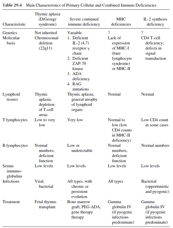
Combined are those in which both cell-mediated immunity and humoral immunity are im-paired. The general characteristics of this group are summarized in Table 29.4.

Severe, Combined Immunodeficiency
Severe, combined immunodeficiency (SCID) is a group of heterogeneous disorders associated with lack of both T- and B-cell function. Most forms are due to an inheritable disorder, which can be inherited as an X-linked recessive form or as an autosomal recessive form.
1. Pathogenesis
There are three predominant pathogenic mechanisms in SCID:
1. Mutation of recombinase-activating genes (RAG) 1, 2, and/or 3, genes that con-trol the V(D)J recombination essential for the differentiation of membrane im-munoglobulins and T-cell receptors. These patients lack both T and B cells, because those two cell populations cannot fully differentiate in the absence of their defining antigen receptors. Those patients are severely im-mune compromised. Inheritance is autosomal recessive.
2. Deficiencies in T-Cell Signaling. The most common form of SCID is X-linked and is due to a mutation in the Îł chain of the IL-2 receptor (IL-2RÎłc or Îłc). This cell surface protein transduces signals delivered by the occupancy of several inter-leukin receptors, including those for IL-2, IL-4, IL-7, IL-11, and IL-15. Mutations in IL-2RÎłc result in absent downstream cell surface signaling through the JAK/STAT signal transduction cascade, a critical component of interleukin-in-duced T-cell activation. A nearly identical SCID phenotype is observed in an au-tosomal recessive defect resulting from a mutation in JAK-3, the downstream pro-tein from IL-2RÎłc. Children with these forms of SCID have no circulating mature CD3+ T cells because of maturation arrest of T-cell development within the thy-mus. B cells are present in normal numbers but are nonfunctional, presumably due to a lack of T-helper cell function. NK cell numbers and function are also deficient.
Zap-70 is a critical intracytoplasmic protein for the transmission of activa-tion signals through the T-cell receptor. Mutations in Zap-70 render this protein nonfunctional, and as a result T-cell activation is blocked. These children’s T cells fail to proliferate in vitro when activated by mi-togens. T-cell enumeration in Zap-70 deficiency reveals normal numbers of CD3+ and CD4+ T cells but low numbers of CD8+ T cells.
3.Deficiencies of purine salvage enzymes. The most common of these disorders are adenosine deaminase (ADA) and purine nucleoside phosphorylase (PNP) de-ficiencies. ADA catabolizes the deamination of adenosine and 2’ -deoxyadeno-sine. Therefore, the lack of ADA causes the intracellular accumulation of these two compounds. 2’ -Deoxyadenosine is phosphorylated intracellularly, and the activity of the phosphorylating enzyme is greater than the activity of the de-phosphorylating enzyme. Consequently, there is a marked accumulation of de-oxyadenosine triphosphate (deoxyATP), which has a feedback inhibition effect on ribonucleotide reductase, an enzyme required for normal DNA synthesis. As a consequence, DNA synthesis will be greatly impaired, and no cell proliferation will be observed after any type of stimulation. In addition, 2’ -deoxyadenosine is reported to cause chromosome breakage, and this mechanism could be the basis for the severe lymphopenia observed in these patients. The reason why lympho-cytes are predominantly affected over other cells that also produce ADA in nor-mal individuals is that immature T cells are among those cells with higher ADA levels (together with brain and gastrointestinal tract cells).
2. Clinical Presentation
In all forms of SCID symptoms start very early in life, usually by 4 months of age. Survival beyond the first year of life is rare without aggressive therapy. Frequent clinical presenta-tions include persistent infections of the lungs, often caused by opportunistic agents such as Pneumocystis carinii, severe mucocutaneous candidiasis, chronic, untractable diarrhea, failure to thrive, and runting. In most cases physical examination shows absence of all lym-phoid tissues: atrophic tonsils, very small or undetectable lymph nodes, signs of pulmonary infection, evidence of poor physical development, and oral thrush. A chest x-ray will reveal absent thymic shadow, bony abnormalities of the ribs in patients with ADA deficiency. Ap-proximately 20% of infants with SCID will develop graft-versus-host disease as a conse-quence of the transplacental transfer of T lymphocytes. The maternal T lymphocytes that enter the fetal circulation are not destroyed because of the infant’s deficient cell-mediated immunity. The maternal T cells can proliferate in the skin, gastrointestinal tract, and liver. Symptoms of graft-versus-host disease include rash, jaundice, hepatitis, and chronic diar-rhea. Severe cases of graft-versus-host disease are often fatal. Children with SCID and other T-cell deficiencies can also develop graft-versus-host disease following blood trans-fusions containing viable mononuclear cells. Children with SCID carry an increased risk of malignancy, particularly lymphomas positive for the Epstein-Barr virus.
3. Diagnosis
The laboratory parameters seen in the various forms of SCID. In most forms these patients have very low lymphocyte counts. In X-linked SCID more than 75% of the residual lymphocyte population are CD19+ B cells, while CD3+ T cells and CD16/56+ NK cells usually represent less than 10% and 2% of the residual population, respectively. The deficiency in cell-mediated immunity is reflected by negative skin tests, delayed rejection of allogeneic skin grafts, and lack of response of cultured mononuclear cells to T-cell mitogens and anti-CD3 monoclonal antibodies. Neutropenia can also be seen in some patients. In cases of ZAP-70 deficiency, lymphocyte counts may be normal or close to normal, but the T lymphocytes do not respond to stimulation.
Immunoglobulins are usually low (levels more than 2SD below the normal level for age) but in some cases can be normal or irregularly affected. B cells and plasma cells are low or undetectable in ADA deficiency and RAG deficiency. In all forms of SCID antibody responses are very low to absent.
The definitive diagnosis and identification of the cause of SCID often requires eval-uation at the molecular level. Many forms of SCID can be diagnosed by molecular analy-sis of the genes or gene products. The diagnosis of ADA deficiency can be made through enzymatic analysis of red blood cells, lymphocytes, fibroblasts, amniotic cells, fetal blood, or chorionic villous samples.
4. Therapy
Children with SCID and other defects in T-cell function should not receive immunizations with live attenuated vaccines. Prophylaxis with trimethoprim-sulfamethoxazole to prevent Pneumocystis carinii pneumonia is indicated. If blood transfusions are necessary, theyshould receive irradiated blood products from donors who test negative for viral infections such as cytomegalovirus. Exposure to all pathogens should be limited, although imple-mentation is not easy (as reflected by the term “bubble baby”).
All forms of SCID can be corrected with a bone marrow transplantation from an HLA-matched sibling. The graft is usually successful, but there is a risk for the develop-ment of GVH disease. If an HLA-matched sibling is not available, alternative strategies include the grafting of haploidentical, T-cell–depleted bone marrow cells from a parent, an HLA-matched unrelated bone marrow donor, or umbilical cord blood leukocytes from an unrelated donor. The risk for GVH disease is greater in any of those modalities. GVH disease is usually prevented with the administration of immunosuppressive drugs follow-ing transplant.
ADA deficiency can be effectively treated by the administration of bovine ADA plus polyethylene glycol (PEG). The addition of PEG results in decreased immunogenicity and increased half-life of the bovine ADA.
It must be noted that ADA deficiency was the first human disease to be successfully treated by gene therapy. The protocol involves harvesting peripheral blood T lymphocytes from the patients, transfecting the ADA gene using a retrovirus vector, expanding the trans-fected cells in culture, and readministering them to the patient. In the first treated patient, normal peripheral blood T-lymphocyte counts and clinical improvement were seen after several such infusions. The infusions need to be periodically repeated, since the ADA+ T-lymphocyte population eventually declines. The normalization of T-cell counts probably reflects the fact that the transfected ADA+ cells will produce excess ADA, which will dif-fuse into genetically deficient cells unable to synthesize it. The therapeutic value of gene therapy in ADA deficiency, however, is limited because all patients have to continue re-ceiving PEG-ADA to maintain a relatively symptom-free status.
Successful gene therapy of SCID secondary to IL-2Rγc deficiency was reported early in 2000. Two children with this form of immunodeficiency received their own CD34+ stem cells after ex vivo transfection of the defective gene by means of a murine retroviral vector. In contrast with ADA gene therapy, the patients with IL2Rγc deficiency have shown long-term immune reconstitution, lasting for 10 months at the time of publica-tion. The recovery of immune functions in these two children was basically complete—T, B, and NK cell counts and function became undistinguishable from those of normal age-matched controls, and so did the capacity of mounting antigen-specific responses.
Related Topics