Chapter: Clinical Cases in Anesthesia : Acute Postoperative Pain
Outline the major afferent pain pathways
Outline the major afferent pain pathways.
Somatic pain is produced by activation of pain
recep-tors in the periphery. There are two principal cutaneous nociceptors:
high-threshold mechanoreceptors (HTMs) and polymodal nociceptors (PMNs). The
HTMs respond to mechanical stimuli and have a receptive field of 1 cm2.
PMNs respond to mechanical, thermal, and chemical irritants. HTM axons conduct
via myelinated A delta fibers at a rate of 5‚Äď25 m/sec and transmit sharp pain
with a rapid onset. PMN axons conduct via unmyelinated C fibers at a rate of
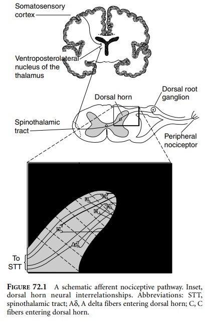
less than 2 m/sec and convey dull aching pain with a slow onset (Figure 72.1).
Pain from visceral origins is carried away from the organs by myelinated
visceral B fibers.

These nerves enter the dorsal horn of the spinal
cord and terminate in different laminae, which were designated histologically
by Rexed. A delta and C fibers synapse with wide dynamic range neurons
(activated by tactile or noxious stimuli) in lamina V and nociceptive-specific
neurons in lamina I. Both types of fibers send branches into the substantia
gelatinosa (laminae II and III), whose inhibitor interneurons modulate
perceived nociception.

The axons of the neurons originating in laminae
V and I cross over to the opposite ventrolateral funiculus of the spinal cord
and ascend as the lateral spinothalamic tract. Some axons terminate in the
ventropostero-lateral nucleus of the thalamus (neospinothalamic tract), where
they synapse with neurons that project to the somatosensory cortex. These
neurons are associated with discrimination (location, duration, and intensity)
of pain. Other axons project to the medial thalamic nuclei as well as the
medulla, pons, midbrain, and hypothalamus (paleo-spinothalamic tract) and are
associated with emotional aspects of pain.
Related Topics