Chapter: Genetics and Molecular Biology: Genes Regulating Development
Outline of Early Drosophila Development
Outline of Early Drosophila Development
As in the development of spores of yeast, a single
diploid cell in the ovary of a female fly gives rise to a number of haploid
cells. In Drosophila the precursor
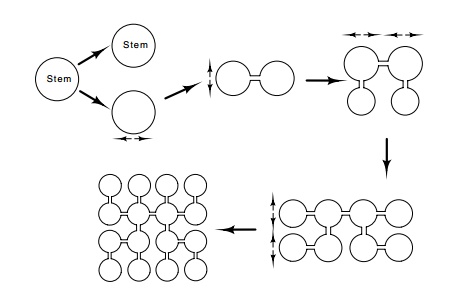
cell undergoes four divisions to yield sixteen cells (Fig. 17.5). These are not
separated, but retain communication with one another via cytoplasmic bridges.
Eventually, one of the two cells with four

Figure
17.5 Creation of nurse and egg cells
from a precursor stem cell in theovary of a Drosophila.
The first division of the stem cell yields one cell which remains stem cell
type and a daughter cell that will yield nurse and egg cells. Four additional
divisions yield a network of interconnected cells.
connections to other cells moves to the end of the
group and begins the development process of generating an egg. The fifteen
connected cells assist the process and are called nurse cells. While it is
developing, the future egg cell is called an oocyte. The nurse cells absorb
nutrients from the surrounding fluid and synthesize rRNA, mRNA, and other
compo-nents. Additionally, maternal diploid cells also influence the developing
oocyte. (Fig. 17.6). These are called the follicle cells, and they surround the
nurse cells and developing oocyte. Partway through the oogenesis pathway, the
nurse cells empty their contents into the developing oocyte. When the
maturation process is complete, the synthetic activi-ties in the follicle cells
almost ceases.
Figure
17.6 Later stage of egg development
showing an egg cell plus nurse cellssurrounded by follicle cells. Although each
cell possesses a nucleus and 16 are present, not all nuclei are in the plane of
the cross section. The future egg cell is at the far right. At a still later
stage of development the egg is enlarging and the nurse cells are found at the
left.

Upon fertilization, DNA synthesis begins at a high rate in the egg. Many replication origins are utilized per chromosome so that a chromosome doubling can take as little as ten minutes.
The original diploid chromosome doubles ten times, then a few nuclei migrate to
the poste-rior pole of the egg where they ultimately become the germ line
cells. The remainder of the nuclei migrate to the surface of the egg and divide
four more times, yielding a total of about 4,000 nuclei. At this point nuclear
membranes and cell walls develop, creating isolated cells. This stage consists
of a single layer of cells and is called a blastula.
Up to the time of wall formation, the nuclei are
not committed to any developmental fate, for they can be moved to different
locations without altering the development of the embryo. After the walls form,
the cells are partially committed, and relocating them interferes with normal
development. About six hours after fertilization the embryo develops about 14
distinguishable segments. Six of these ultimately fuse to yield the head, three
become the thorax, and about eight develop into the abdomen. By 24 hours after
fertilization the larva is fully developed.
Related Topics