Chapter: Medical Physiology: The Eye: I. Optics of Vision
Ophthalmoscope

Ophthalmoscope
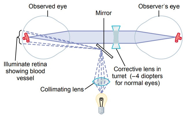
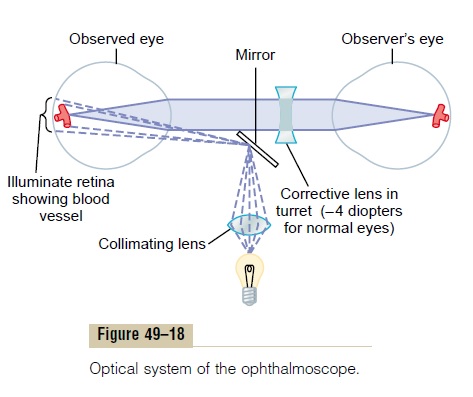
The ophthalmoscope is an instrument through which an observer can look into another person’s eye and see the retina with clarity. Although the ophthalmoscope appears to be a relatively complicated instrument, its principles are simple. The basic components are shown in Figure 49–18 and can be explained as follows.

If a bright spot of light is on the retina of an emmetropic eye, light rays from this spot diverge toward the lens system of the eye. After passing through the lens system, they are parallel with one another because the retina is located one focal length distance behind the lens system. Then, when these parallel rays pass into an emmetropic eye of another person, they focus again to a point focus on the retina of the second person, because his or her retina is also one focal length dis-tance behind the lens. Any spot of light on the retina of the observed eye projects to a focal spot on the retina of the observing eye. Thus, if the retina of one person is made to emit light, the image of his or her retina will be focused on the retina of the observer, provided the two eyes are emmetropic and are simply looking into each other.
To make an ophthalmoscope, one need only devise a means for illuminating the retina to be examined. Then, the reflected light from that retina can be seen by the observer simply by putting the two eyes close to each other. To illuminate the retina of the observed eye, an angulated mirror or a segment of a prism is placed in front of the observed eye in such a manner, as shown in Figure 49–18, that light from a bulb is reflected into the observed eye. Thus, the retina is illuminated through the pupil, and the observer sees into the subject’s pupil by looking over the edge of the mirror or prism or through an appropriately designed prism.
It is clear that these principles apply only to people with completely emmetropic eyes. If the refractive power of either the observed eye or the observer’s eye is abnormal, it is necessary to correct the refractive power for the observer to see a sharp image of the observed retina. The usual ophthalmoscope has a series of very small lenses mounted on a turret so that the turret can be rotated from one lens to another until the correction for abnormal refraction is made by selecting a lens of appropriate strength. In normal young adults, natural accommodative reflexes occur that cause an approximate +2-diopter increase in strength of the lens of each eye. To correct for this, it is necessary that the lens turret be rotated to approximately –4-diopter correction.
Related Topics