Chapter: Medical Physiology: Excitation of Skeletal Muscle: Neuromuscular Transmission and Excitation-Contraction Coupling
Muscle Action Potential
Muscle Action Potential
Some of the quantitative aspects of muscle potentials are the following:
1. Resting membrane potential: about –80 to –90 millivolts in skeletal fibers—the same as in large myelinated nerve fibers.
2. Duration of action potential: 1 to 5 milliseconds in skeletal muscle—about five times as long as in large myelinated nerves.
3. Velocity of conduction: 3 to 5 m/sec—about 1/13 the velocity of conduction in the large myelinated nerve fibers that excite skeletal muscle.
Spread of the Action Potential to the Interior of the Muscle Fiber by Way of “Transverse Tubules”
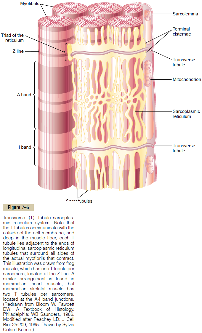
The skeletal muscle fiber is so large that action poten-tials spreading along its surface membrane cause almost no current flow deep within the fiber. Yet, to cause maximum muscle contraction, current must pen-etrate deeply into the muscle fiber to the vicinity of the separate myofibrils. This is achieved by transmission of action potentials along transverse tubules (T tubules) that penetrate all the way through the muscle fiber from one side of the fiber to the other, as illustrated in Figure 7–5. The T tubule action potentials cause release of calcium ions inside the muscle fiber in the immediate vicinity of the myofibrils, and these calcium ions then cause contraction. This overall process is called excitation-contraction coupling.

Related Topics