Chapter: Anatomy of Flowering Plants: An Introduction to Structure and Development : Leaf
Leaf Vasculature
Leaf Vasculature
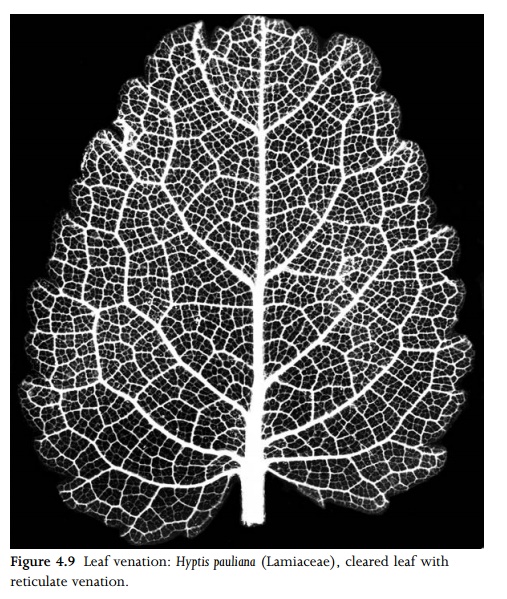
There are two main leaf venation types among the angiosperms: parallel and reticulate. Broadly, parallel venation is typical of mono-cots and reticulate venation of eudicots and magnoliids, though there are many exceptions. In leaves with parallel venation the main veins (primary veins) are parallel for most of their length and converge or fuse at the leaf tip. Typically, numerous small veins interconnect the larger veins, but there are very few vein endings
in the
mesophyll. In leaves with reticulate venation (Fig. 4.9) there is often a major
vein in the middle of the leaf, the midrib or primary vein, which is continuous
with the major venation of the petiole. The midrib is linked to many smaller
secondary (second-order) veins that branch from it and often extend to the leaf
margins. Secondary veins sometimes terminate in a hydathode at the leaf margin.
In their turn, smaller veins branch from the second- and subsequent-order
venation, forming a reticulate network. The areas of mesophyll between the
smallest veins in the leaf are termed areoles. In many species small veins
branch into the areoles to form vein endings. Variable aspects of leaf venation
include the relative number of veinlet endings per areole, and whether
second-order veins terminate at the margins or loop around to link with the
superadjacent secondary veins.
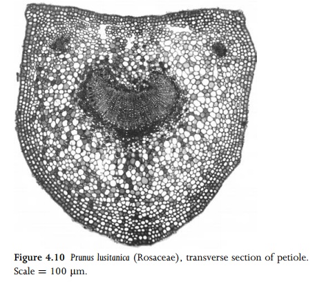
Petioles also possess characteristic venation. The simplest form of petiole vasculature appears in transverse section as a crescent with
xylem on the adaxial side and phloem on the abaxial side (Fig. 4.10). Some
species possess additional bundles outside the main vascular crescent, which
may itself be inrolled at the ends, or in a ring, or divided into separate
bundles. Classification of the various forms of petiole vasculature depends on
how it is linked to the stem vasculature at the node. One or more vascular
traces may depart from each gap in the stem vascular cylinder. The number and
pattern of vascular bundles sometimes vary along the length of the petiole.
Midrib vasculature, which is continuous with that of the petiole, is subject to
similar variation.
In
transverse sections of the lamina, vascular bundles are usually arranged in a
single row. However, in some species with very thick leaves, such as Agave,
there are two or more rows of vascular bundles. Lamina bundles are usually
collateral, with adaxial xylem and abaxial phloem, but orientation can vary,
and in some cases bundles are bicollateral or even amphivasal. In the
isobilateral leaves of some monocots there are two rows of vascular bundles
with opposite orientation to each other, the xylem poles being oriented towards
the leaf centre. Centric leaves possess a ring of vascular bundles.
Leaf
vasculature develops acropetally from the primordial pro-cambial strand at its
base. The central trace develops first, and ultimately becomes the
midvein. The xylem conducting system of the leaf blade often consists entirely
of tracheids, usually with helical or annular thickenings, though in some
leaves both vessel elements and xylem parenchyma are also present. The smallest
vascular bundles often consist of only one or two rows of xylem tracheids and a
few files of phloem sieve tube elements.
Related Topics