Chapter: Maternal and Child Health Nursing : Fetal Development, Placenta Development and Fetal Circulation
Growth and Development of the Fertilised Ovum
Growth and Development of the
Fertilised Ovum
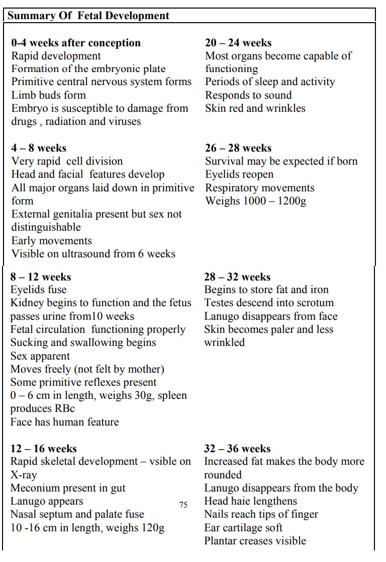
During
the first 8 weeks of pregnancy, embryonic tissues and the surrounding
supportive structures are formed simultaneously. It is during this period that
the embryo is at greatest risk for malformation. From the 8th week
through the end of pregnancy, the embryo is known as the FETUS. The supportive
structures that nourish and maintain the growing fetus are called the fetal
membranes. These include the yolk sac, amnion, chorion, decidua and the
placenta.
The Trophoblast
As
development continues small projections begin to appear all over the surface of
the blastocyst known as the tropoblast, becoming most prolific at the area of
contact – are a of inner cell mass. The trophoblast differentiates into layers.
1.
The outer syncytiotropoblast (syncitium): it is
capable of breaking the decidua tissue during embedding. It erodes the wall of
the blood vessels, making nutrient in the maternal blood accessible to the
developing embryo. It acts as a protective layer between the chorionic villi.
2.
Cytotrophoblast: This is a well defined single
layer of cells which produce Human Chorionic gonadotrophin (HCG). It informs
the corpus Luteum that pregnancy has begun, so as to continue to produce
progesterone and oestrogen. The progesterone maintain the integrity of the
decidua so that shedding does not take place (menstruation is suppressed),
while the high level of oestrogen suppresses the production of FSH. The HCG is
produced in high level in the first trimester and it is the basis for pregnancy
test.
3.
The Mesoderm: Consist of loose connective tissue.
It is continuous with that in the inner cell mass where they join in the body
stalk which later develops into the umbilical cord.
The
trophoblast later form finger like process called –Primitive villi which
develop into placenta and the chorion.
The Inner Cell Mass
As the
trophoblast is developing into the placenta which will nourish the fetus, the
inner cell mass is forming the fetus itself, umbilical cord and the amnion. The
cells differentiate into three layers each of which will form particular parts
of the fetus.
The Ectoderm: Mainly forms the skin, nervous
system,mammary glands salivary glands, Pharynx, nasal passage and crystalline
lens of the eyes, certain lining of the mucosa, hair, nails, and enamel of the
teeth.
The Mesoderm: Forms the bones muscles,
circulatory system oldvessels Reproductory system (ovary and testes), kidneys,
ureters, connective tissues, lymphatic system.
The Endoderm: Lines the yolk sac. It forms the
Alimentary tract,liver, pancreas, lungs, Bladder thyroid glands.
The fetus
develops it’s own blood like other organs in the body. The maternal and the
fetal blood never mix. During the later weeks (4 wks) the organs like the liver
and heart start to function.
The three
layers together are known as the embryonic
plate. Two cavities appear in the inner cell mass one on either sides of
the embryonic plate.
1. The Amniotic Cavity: this lies
on the side of theectoderm. The cavity which is filled with fluid gradually
enlarges and fold round the embryo to enclose it the lining forms the amnion.
It later enlarges in the chronic cavity and comes in contact with the chorionic
membrane.
2. The Yolk
Sac: Lies on the side of the endoderm andprovides nourishment for the embryo
until the placenta(alimentary tract
After
birth the remnants of the yolk sac is the vestigial structure in the base of
the umbilical cord, known as vitelline duct.
The
developing of spring is referred as EMBRYO after fertilization up to 8 weeks
after which the conceptus is known as FETUS until birth.
Related Topics