Chapter: Obstetric and Gynecological Nursing : Anatomy of Female Pelvis and the Fetal Skull
Female Pelvic Bones
Female Pelvic Bones
The female pelvis is structurally adapted for child beaing and delivery.
There are four pelvic bones
·
innominate or hip bones
·
Sacrum
·
Coccyx
A. Innominate
bones
Each innominate bone is composed of three parts.
1.
The ilium the large flared out part
2.
The ischium the thick lower part. It has a large prominance known as the
ischial tuberosity on which the body rests when sitting. Behind and a little
above the tuberosity is an inward projection, the ischial spine. In labour the
station of the fetal head is estimated in relation to ischial spines.
3.
The pubis - The pubic bone forms the anterior part.
The space enclosed by the body of the pubic bone the rami and the
ischium is called the obturator foramen.
B. The sacrum - awedge shaped bone consisting of five fused vertebrae.
The upper border of the first sacral vertebra is known as the sacral
promontary. The anterior surface of the sacrum is concave and is referred to as
the hallow of the sacrum.
C. The coccyx: - is avestigial tail. It consists of four fused vertebrae
forming a small triangular bone.
Pelvic Joints
There are four pelvic joints
·
One Symphysis pubis
·
Two Sacro illiac joint
·
One Sacro coccygeal joint
·
The symphysis pubis is a cartilgeous joint formed by junction of the two
pubic bones along the midline.
·
The sacro iliac joints are the strongest joints in the body.
·
The sacro coccygeal joint is formed where the base of the coccyx
articulates with the tip of the sacrum.
In non pregnant state there is very little movement in these joints but
during pregnancy endocrine activity causes the ligaments to soften which allows
the joints to give & provide more room for the fetal head as it passes
through the pelvis.
Pelvic ligaments
·
Interpubic ligaments at the symphysis pubis (1)
·
Sacro iliac ligaments (2)
·
Sacro coccygeal ligaments (1)
·
Sacro tuberous ligament (2)
·
Sacro spinous ligament (2)
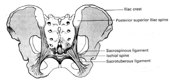
The True Pelvis
The true pelvis is the bony canal through which the fetus must pass
during birth. It has a brim, mid cavity and an out let. The pelvic brim is
rounded except where the sacral promontory projects into it. The pelvic cavity
is extends from the brim above to the out let below. The pelvic out let are two
and described as the anatomical and the obstetrical. The anatomical out let is
formed by the lower borders of each of the bones together with the
sacrotuberous ligament. It is diamond in shape. The obstretrical out let is of
the space between the narrow pelvic strait and the anatomical outlet.
Important land marks of female pelvis
A. Pelvic brim
·
Sacral promentary posteriorly
·
Superior ramus of the pubic bone antro lateral
·
Upper inner boarder of the body of the pubic bone
·
Upper inner boarder of the symphysis pubis anteriorly
B. Mid pelvis
·
Ischial spine
C. Out let
·
Inferior pubic rami antero laterally
·
Sacrotuberous ligament postro laterally
·
Ischial tuberosity laterally
·
Inferior border of symphsis pubis anteriorly.
·
Tip of coccyx
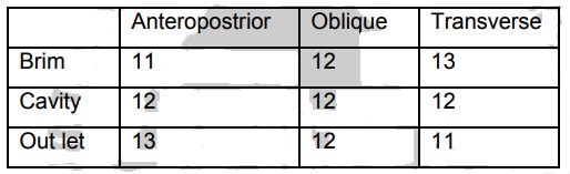
Important diameters of the pelvis
Inlet
Diagonal conjugate - a line from the sacral promontory toward the lower
boarder of the symphysis pubis and measures 12.5 centimeter. It is measured by
pelvic examination.
Mid cavity
Interspinous diameter-a line between the two ischial spines and measures
11 centimeter.
The pelvic out let
·
Pubic arch
· Intertuberous diameter
Table 1. Measurements of the pelvic canal in centimeters
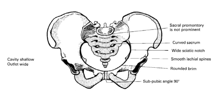
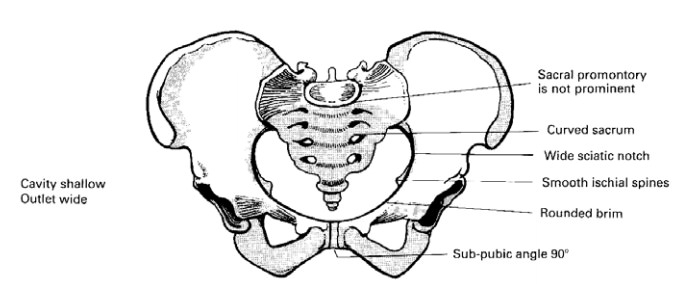
The four types of female pelvis
·
The gynacoid pelvis (female type)
·
The android pelvis (male type)
·
The anthropoid pelvis
·
The platypelloid pelvis
Table 2 .Features of the four types of female pelvis
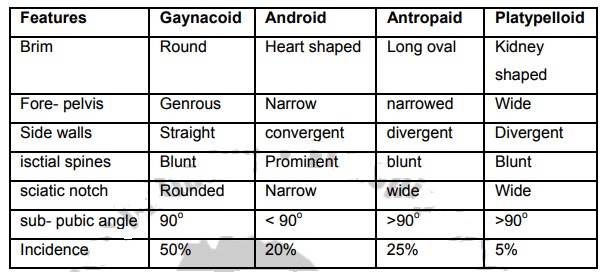
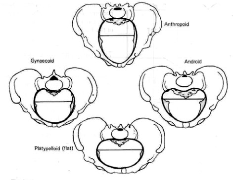
Figure 3 Types of female pelvis (Alan H. Decherney l. pemoll,
1994)
Pelvic floor Or Pelvic diaphragm
The pelvic floor or diaphragm is amuscular floor that demarcates the
pelvic cavity and perineum. Its strength is inforced by its associated condesed
pelvic fascia, therefore, it is important for pelvic organs protection.
Functions: -
It supports the weight of the abdominal and pelvic organs
The muscles are responssible for the voluntary control of micturation,
defication and play an important part in sexual intercourse.
It infulences the passive movement of the fetus through the birth canal
and relaxes to allow its exit from the pelvis.
The main important muscels of pelivic floor are:
·
Levater ani muscles are arising from the lateral pelveic wall and
decussate in the midline between the urethra, the Vagina and rectum. It
contains pubococcygeous muscle, ileo coccygeus and pubo rectalis.
· Pubococygeous muscle is constructed in such away that it can expand enough for child bith and contract the pelvis supported
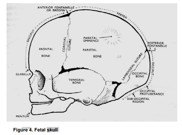
The Fetal Skull
The fetal head is the most difficult part to deliver whether it comes
first or last. It is large in comparison with the ture pelvis and some
adptation between skull and pelvis must take place during labour.An
understanding of the landmarks and measurements of the fetal skull enables to
recognize normal presentation and positions and to facilitate delivery with the
least possible trauma to mother and child. The skull is divided into the vault,
the base and the face. The vault is the large dome shaped part above the
imaginary line drowns between the orbital ridges and the nape of the neck.
The base is composed of bones which are firmly united to protect the
vital centres in the medulla.
The face is composed of 14 small bones which are also firmly united and
non- compressible
Bones of the Vault
There are five main bones in the vault of the fetal skull.
1.
The occipital bone lies at the back of the head and forms the region of
the occiput.
2.
The two parietal bones lie on either side of the skull.
3. The two frontal bones from the forehead or sinciput.
Sutures and fontanelles
Sutures are cranial joints and are formed where two bones adjoin. Where
two or more sutures meet, a fontanell is formed.
Types of sutures
1.
The lambdoidal suture is shped like the Greek letter lambda and
separates the occipital bone from the two parital bones.
2.
The sagital suture lies between the parital bones
3.
The coronal sutrue separetes the frontal bones from the parital bones,
passing from one temple to the other.
4.
The frontal suture runs between the two haves of the frontal bone
Types of fontanelle
1.
The posterior fontanelle or lambda is situated at the junction of the
lambdiodal and sagital sutures. It is small triangular in shape and can be
recogonized vaginally.
2.
The anterior fontanelle or bregma is found at the junction of the
sagital, coronal and frontal sutures and recognized vaginally.
The sutures and fontanelles, because they consist of memberanous spaces,
allow for a degree of overlapping of the skull bones during labour and
delivery.
Regions of the Skull
ii. The vertex is bounded by the posterior fontanelle, the parital
eminences and the anterior fontanelle. Of the 96% of the babies born head
first, 95% present by the vertex.
iii. The sinciput or brow extends from the anterior fontanelle and the
coronal suture to the orbital ridges.
iv. The face is small in new
born baby. It extends from the orbital ridges and the root of the nose to the
junctions of the chin and the neck. The point between the eye brows is knowns
as the glabella. The chin termed the mentum and is an important land mark.
Land Marks of the Fetal Skull
i. Sinciput
ii. Occiput
iii. Glabella
iv. Anterior fontanelle
v. The vertex
vi. Posterior fontanelle
vii. Occuputal protuberunse
viii. The mentum
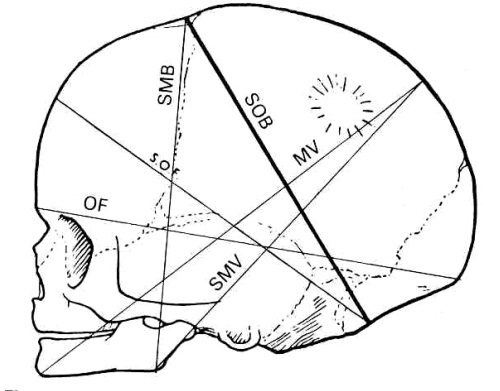
Diameters of the Fetal Skull
The measurement of the skulls are transverse,
anteropositerior or longitudinal.
• Transverse diametes
. Biparietal diameter 9.5 cm between the parietaleminence
. Bitemporal diameter 8.2cm between the furthersepoints of
the coronal suture at the temples.
• Anteroposterior
or longitudinal diameters
. Suboccipitobregmatic 9.5 cm from below the
occipitalprotuberance to the center of the anterior fontanelle or bregma
. Suboccipitofrontal
10cm from below occipital protuberance to the center of the frontal suture.
. Occipitofrontal
11.5 cm from the occipital protuberance to the glabella.
. Mentovertical 13.5cm from the point of the chin to the highest point on the vertex sightly nearer to the posterior than to the anterior fontanelle.
. Submentovertical
11.5 cm from the point where the chin joins the neck to the highest point on
the vertex.
. Submentobregmatic
9.5cm from the point where the chin joins the neck to the center of the bregma.
Related Topics