Chapter: Obstetric and Gynecological Nursing : Anatomy of Female Pelvis and the Fetal Skull
The Breast Anatomy

The Breast Anatomy
The female breasts
The female breasts, also known as the mammary glands, are accessory
orgns of reproduction.
Situation One breast is situated on each
side of the sternumand extends between the levels of the second and sixth rib.
The breasts lie in the superficial fascia of the chest wall over the pectoralis
major muscle, and are stabilized by suspensory ligaments.
Shape Each breast is a hemispherical
swelling and has a tailof tissue extending towards the axilla (the axillary
tail of spence).
Size The size varies with each
individual and with the stage ofdevelopment as well as with age. It is not
uncommon for one breast to be little or larger than the other.
Gross structure
The axillary tail is the breast tissue extending
towards theaxilla.
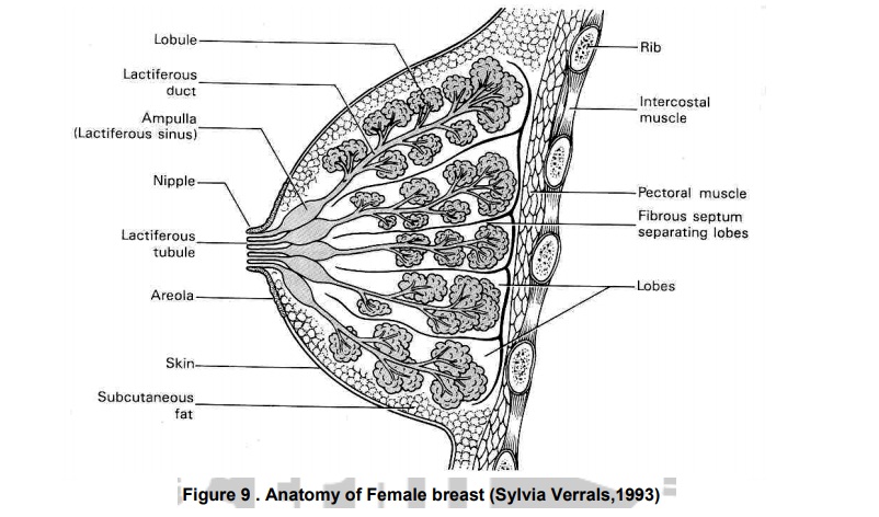
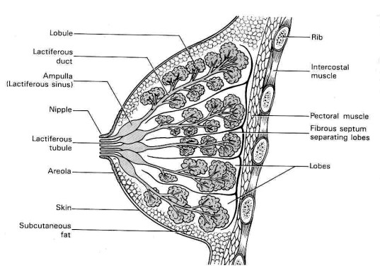
The areoa is a circular area of loose,
pigmented skin about2.5 cm in diameter the centre of each breast. It is a pale
pink colour in a fair- skinned woman, darker in a brunett, the colour deepening
with pregnancy. Within the area of the areola lie approximately 20 sebaceous glands.
In pregnancy these enlarge and are known as montgeomery’s tubercles.
The nipple lies in the centre of the areola
at the level of thefourth rib. Aprotuberance about 6mm in length, composed of
pigmented erectile tissue.The surface of the nipple is perforarted by small
orifices which are the openings of the lactiferous ducts. It is covered with
epithelium.
Microscopic structure The breast is composed largely
ofglandular tissue, but also of some fatty tissue, and is covered with skin.
This glandular tissue is divided into about 18 lobes which are completely
separated by bands of fibrous tissue. The internal structure is said to be
resemble as the segments of a halved grape fruit or orgnge. Each lobe is a
self-contained working unit and is composed of the following structures
Alveoli: Containing the milk- secreting
cells. Each alveolus islined by millk-secreting cells, the acini, which extract from the mammary blood
supply the factors essential for milk formation. Around each alveolus lie
myoepithelial cells, sometimes called ‘basket’ or ‘spider’s cells. When these
cells are stimulated by oxytocin they contract releasing milk into the
lactifierous duct.
Lactifierous tubules:small
ducts which connect the alveoli.
Lactifierous duct:a
central duct into which the tubules run.
Amplulla:the
widened-out portion of the duct where milk isstored. The ampullae lie under the
areola.
Blood supply Blood is supplied to the breast
by the internalmammary, the external mammary and the upper intercostals
arteries.Venous drainage is through corresponding vessles into the internal
mammary and axillary veins.
Lymphatic drainage This is largely into the
axillary glands, with some dranage
in to the portal fissure of the liver and mediastinal glands. The lymphatic
vessels of each breast communicate with one another.
Nerve supply The function of the breast is
largely controlledby hormone activity but the skin is supplied by breanches of
the thoracic nerves. There is also some sympathetic nerve supply, especially
around the areola and nipple.

Related Topics