Chapter: Medical Microbiology: An Introduction to Infectious Diseases: Enteroviruses
Enteroviruses: Group Characteristics - Virology
ENTEROVIRUSES: GROUP CHARACTERISTICS
VIROLOGY
MORPHOLOGY AND BIOLOGICAL FEATURES
As a group, the enteroviruses are extremely small (22 to 30 nm in diameter), naked viri-ons with icosahedral symmetry. They possess single-stranded, positive-sense RNA and a capsid formed from 60 copies of four nonglycosylated proteins (VP1, VP2, VP3, VP4). Replication and assembly occurs exclusively in the cellular cytoplasm; one infectious cy-cle can occur within 6 to 7 hours. This results in cessation of host cell protein synthesis and cell lysis with release of new infectious progeny.
Unlike rhinoviruses, which are also members of the picornavirus family, enteroviruses are quite resistant to an acid pH (as low as 3.0). This feature undoubtedly helps ensure their survival during passage through the stomach to the intestines. Enteroviruses are also resistant to many common disinfectants such as 70% alcohol, substituted phenolics, ether, and various detergents that readily inactivate most enveloped viruses. Chemical agents, such as 0.3% formaldehyde or free residual chlorine at 0.3 to 0.5 ppm, are effective; how-ever, if sufficient extraneous organic debris is present, the virus can be protected and sur-vive long periods.
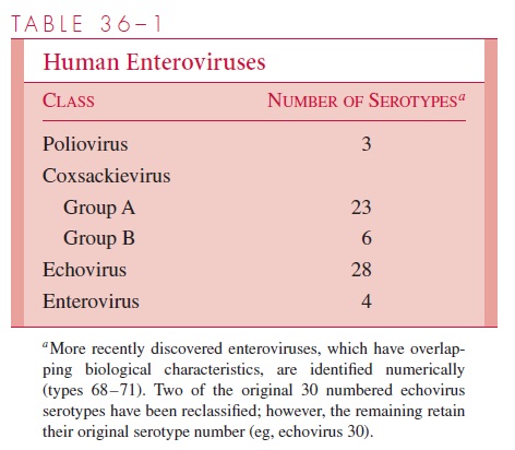
Some of the enterovirus serotypes share common antigens, but there are no significant serologic relationships between the major classes listed in Table 36–1. Genetic variation within specific strains occurs, and mutants that exhibit antigenic drift and altered tropism for specific cell types are now recognized. Polioviruses, which have been most extensively studied as enterovirus prototypes, are known to have epitopes on three surface structural proteins (VP1, VP2, and VP3) that induce type-specific neutralizing antibodies. This ap-pears to be generally the case for all enteroviruses; definitive identification of isolates usu-ally requires neutralization tests.
GROWTH IN THE LABORATORY
Most enteroviruses can be isolated in primate (human or simian) cell cultures and show characteristic cytopathic effects. Some strains, particularly several coxsackievirus A serotypes, are more readily detected by inoculation of newborn mice. In fact, the new-born mouse is one basis for originally classifying group A and B coxsackieviruses. Group A coxsackieviruses cause primarily a widespread, inflammatory, necrotic effect on skeletal muscle, leading to flaccid paralysis and death. Similar inoculation of group B coxsackieviruses causes encephalitis, resulting in spasticity and occasionally convul-sions. Echoviruses and polioviruses rarely have an adverse effect on mice, unless spe-cial adaptation procedures are first employed. The higher-numbered enteroviruses (types 68–71), which have overlapping, variable growth and host characteristics, have been classified separately.
Related Topics