Chapter: 12th Microbiology : Chapter 5 : Food Microbiology
Diary Microbiology
Diary Microbiology
The area
of dairy microbiology is large and diverse. The bacteria in dairy products may
cause disease or spoilage. Some bacteria may be specifically added to milk for
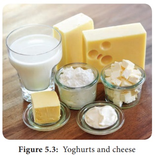
fermentation to produce products like yoghurts and cheese (Figure 5.3).
MILK
Milk is
the fluid, secreted by mammals for the nourishment of their young ones. It is
in liquid form without having any colostrum. The milk contains water, fat,
protein and lactose. About 80–85% of the protein is casein. Due to moderate pH
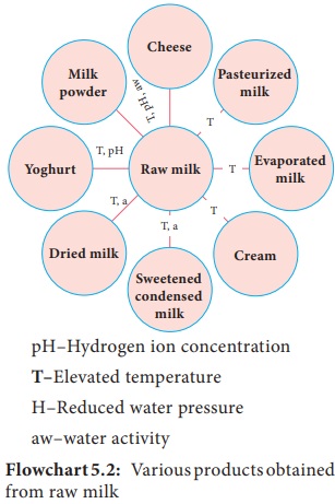
(6.4–6.6), good quantity of nutrients and high water content, milk an excellent
nutrient for the microbial growth. (Flowchart 5.2).
Composition and Properties
Milk is
considered to be the “Most nearly perfect” food for man and hence is one of the
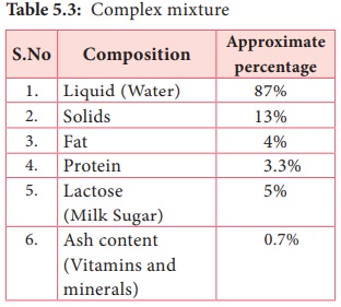
most important ingredients of the diet. It is an extremely complex mixture and
usually contains (Table 5.3).
pH–Hydrogen
ion concentration
T–Elevated temperature
H–Reduced
water pressure
aw–water
activity
Sources of Microorganisms in Milk
• Three
sources contribute to the microorganism
found in milk the udder interior, the teat exterior and its immediate
surroundings, and the milking and milk handling equipment.
• Bacteria that get on to the outside of the teat may be able to invade the opening and hence the udder interior. The organisms most commonly isolated are Micrococcus, Streptococci and the diptheroid Corynebacterium bovis. Aseptically taken milk from a healthy cow normally contains low number of organisms, typically fewer than 102–103 cfu ml-1
• The
udder exterior and its immediate environment
can be contaminated with organisms from the cow’s general environment. Heavily
contaminated teats have been reported to contribute up to 105 cfu ml-1 in the
milk. Contamination from bedding and manure can be source of human pathogens
such as E.Coli, Campylobacter, Salmonella, Bacillus spp. and Clostridia spp.
• Milk – handling equipment such as teat cups, pipe work, milk holders and storage tanks is the principal source of the microorganisms found in raw milk. Micrococcus and Enterococcus.
Microbiological Standard and Grading of Milk
In India,
raw milk is graded by Bureau of Indian standards (BIS) 1977. The Indian
standard institute (ISI) has prescribed microbiological standard for quality of
milk.
• Coliforms
count in raw milk is satisfactory
if, coliforms are absent in 1:100 dilution.
• Coliforms
count in pasteurized milk is satisfactory
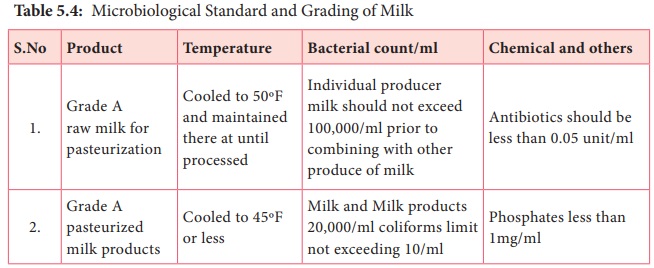
is coliforms are absent in 1: 10 dilution (Table 5.4)
Grading of milk
The quality of milk is judged by certain standards and it is known as grading milk. Grading of milk is based upon regulations pertaining to production, processing and distribution. This includes sanitation, pasteurization, holding conditions and microbiological standards. The U.S public health secrine publication “Milk ordinance and code” shows the following chemical, bacteriological and temperature standards for grade A milk and milk products.
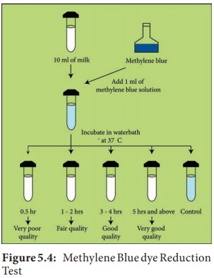
Methylene Blue dye Reduction Test (MBRT)
Methylene
blue dye reduction test commonly known as MBRT test is used as a quick method
to access the microbiological quality of raw and pasteurized milk. This test is
based on the fact that the blue colour of the dye solution added to the milk
get decolorized when the oxygen present in the milk get exhausted due to
microbial activity. The sooner the de colorization, more inferior is the
bacteriological quality of milk assumed to be MBRT test may be utilized for
grading of milk which may be useful for the milk processor to take a decision
on further processing of milk.
Procedure
The test
has to be done under sterile conditions. Take 10ml milk sample in sterile MBRT
test tube. Add 1 ml Methylene Blue dye solution (dye concentration 0.005%).
Stopper the tubes with sterilized rubber stopper and carefully place them in a
test tube stand dipped in a serological water bath maintained at 37°C, records
this time as the beginning of the incubation period. Decolourization is
considered complete when only a faint blue ring (about 5mm) persists at the top
(Figure 5.4).
Recording
of Results – During incubation, observe colour changes as follows:
a. If any
sample is decolourized on incubation for 30 minutes, record the reduction time
as MBRT 30 minutes.
b. Record
such readings as, reduction times in whole hours. For example, if the colour
disappears between 0.5 and 1.5 hour readings, record the result as MBRT 1 hour,
similarly, if between 1.5 and 2.5 hours as MBRT-2 hour and so on.
c.
Immediately after each, reading, remove and record all the decolourized samples
and then gently invert the remaining tubes if the decolourization has not yet
begun (Table 5.5).

Related Topics