Chapter: Human Neuroanatomy(Fundamental and Clinical): Further Consideration of the Cerebral Cortex
Motor and Premotor Areas of the Cerebral Cortex
Motor and Premotor Areas
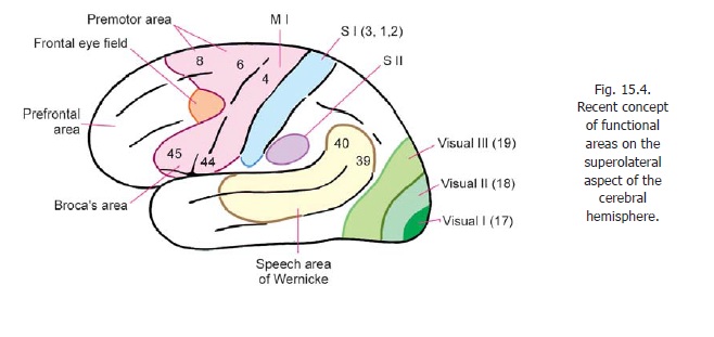
As described the motor area lies in the precentral gyrus (area 4). This is theprimarymotor area (MI). This area has the lowest threshold of stimulation for producing contralateral bodymovements.
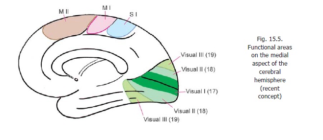
A supplemental motor area (MII) is present on the medial aspect of the cerebral hemisphere, in the medial frontal gyrus, anterior to the paracentral lobule (areas 6, 8). In this area the body is represented with the area for the lower limb posteriorly, that for the face anteriorly, the area for the upper limb lying between these two. Additional motor areas have been described in the adjoining part of the cingulate gyrus.
On the superolateral surface, the premotor area lies anterior to the motor area occupying areas 6 and 8. (We have seen that on the medial aspect of the hemisphere areas 6 and 8 are occupied by the supplemental motor area.)
Advanced:
The distinction between motor and premotor areas is vague, and the entire area is sometimes described as the primary motor area. Studies on areas of the cortex giving origin to corticospinal fibres show that they arise from a wide area covering the main and supplemental motor areas, the premotor area, and many areas in the parietal lobe (SI including areas 3, 2, 1; area 5; and SII in the parietal operculum). It is obvious that this area is not coextensive with the definition of motor areas. Some studies claim that only 20 - 30% of corticospinal fibres arise from area 4; and that as many as 50% may have their origin in the parietal lobe. In relation to motor function the site of termination of corticospinal fibres in spinal grey matter becomes significant. Most fibres that descend from the parietal lobe end in the dorsal grey column; while those ending in the ventral grey column are predominantly from the frontal lobe. The latter appear to be the ones most important for motor control.
The premotor area appears to be responsible for programming intended movements, and control of movements in progress. It is divisible into a dorsal and a ventral area. The dorsal area appears to be concerned with movements initiated by the individual, while the ventral area appears to be concerned with control of movements that take place in response to external stimulation.
The cortex of the motor area is characterised by the presence of large pyramidal cells (giant pyramidal cells of Betz). At one time these cells were considered to be the source of all corticospinal fibres but, as discussed above, this if now known to be incorrect.
It has been estimated that the size of the soma of a neuron is proportional to the total volume of its processes. It follows that neurons having very long axons will have large somata. Purkinje cells that give origin to corticospinal fibres descending to sacral segments of the spinal cord can, therefore, be expected to be among the largest pyramidal cells.
Apart from its corticospinal output, the motor area is connected (in a point to point manner) with the main sensory cortex (SI). This explains why neurons in area 4 are responsive to peripheral stimulation. Area 4 receives afferents from the posterior part of the ventral lateral nucleus of the thalamus. This nucleus receives fibres from cerebellar nuclei. In this way the cerebellum projects to area 4. Area 4 also receives fibres from some other parts of the thalamus, the hypothalamus, and some other parts of the cerebral cortex (including the primary sensory area SI).
The frontal eye field and the motor speech area (of Broca) are parts of the premotor area.
Apart from the motor speech area of Broca there are two other areas concerned with motor control of speech. One of these is located in the temporal and parietal lobes (motor speech area of Wernicke) while the other is located in the supplemental motor area (MII) (Figs. 15.4, 15.5).
Clinical:
In summary note the following:
a) A localised lesion of the primary motor area normally produces contralateral monoplegia. An extensive lesion can cause hemiplegia.
b) Lesions of the premotor area have an adverse effect on skilled movements.
c) Lesions of the frontal eye field result in deviation of both eyes to the side of lesion.
d) Lesions of the motor speech area destroy the ability to speak.
e) Lesions of the prefrontal areas lead to personality changes.
Related Topics